Medical Care |

Medical Care

##SEVER##

/h/herzzentrum-basel.ch1.html


Herzzentrum-basel.ch

Rapid Dynamics of Polyomavirus Type BKin Renal Transplant Recipients Georg A. Funk,1,2 Ju¨rg Steiger,3 and Hans H. Hirsch2,4
1School of Biological Sciences, Royal Holloway University of London, Egham, United Kingdom; 2Transplantation Virology, Medical Microbiology,Department of Clinical and Biological Sciences, University of Basel, and 3Transplantation Immunology and Nephrology and 4Infectious Diseasesand Hospital Epidemiology, University Hospitals Basel, Basel, Switzerland Polyomavirus type BK–associated nephropathy (PVAN) is an emerging cause of early renal transplant failure. No specific antiviral treatment has been established. Current interventions rely on improving immune functionsby reducing immunosuppression. In patients with PVAN, a high BK virus (BKV) load is detectable in plasma. However,the relationship between BKV replication and disease is not well understood.
In a retrospective analysis of BKV plasma load in renal transplant recipients undergoing allograft nephrectomy (n p 3) or changes in immunosuppressive regimen (n p 12), we calculated viral clearance rates andgeneration times and estimated the loss of BKV-infected renal cells.
After nephrectomy, BKV clearance was fast (viral half-life [t ], 1–2 h) or moderately fast (t , 20– 38 h), depending on the sampling density, but it was independent of continued immunosuppressive regimens.
After changing immunosuppressive regimens, BKV was cleared with a t of 6 h–17 days. Using the basic repro- ductive ratio, the efficacies of intervention ranged from 7% to 83% (mean, 28%; median, 22%).
The results emphasize that high-level BKV replication is a major pathogenetic factor that may have implications for genome rearrangements, immune evasion, and antiviral resistance.
Polyomavirus type BK–associated nephropathy (PVAN) load has been detected in the plasma of renal transplant affects 1%–10% of renal transplant recipients, with al- recipients [3, 8]. Interestingly, BKV plasma virus load lograft failure as high as 80% [1–5]. Although PVAN has been reported to rapidly disappear after the surgical likely results from multiple, partly complementary de- removal of renal allografts, regardless of the continu- terminants [5, 6], intense immunosuppression is gen- ation of immunosuppressive regimens [5]. A decrease erally accepted as being the major risk factor [7]. Be- in BKV plasma load was also observed after a reduction cause specific antiviral treatments are not available, in immunosuppressive regimens [3]. Thus, BKV virus reducing immunosuppressive regimens is the current load has been proposed as surrogate marker of PVAN, mainstay of intervention [7]. In the absence of inter- to guide diagnosis and the treatment response. How- vention, PVAN relentlessly progresses to irreversible al- ever, the relationship between BKV replication and dis- lograft failure with extensive fibrosis and tubular at- ease is not well understood [9]. In a retrospective anal- rophy. Throughout these stages, a high BK virus (BKV) ysis, we used mathematical modeling to analyze the BKVplasma load observed in patients after allograft nephrec-tomy and compared the results with those obtained afterchanges in immunosuppressive regimens.
Received 5 April 2005; accepted 18 July 2005; electronically published 30 November 2005.
Potential conflicts of interest: none reported.
Financial support: European Community (Marie Curie Fellowship, contract MEIF- CT-2004-501039 to G.A.F.); Ausbildungsstiftung fu¨r den Kanton Schwyz und dieBezirke See und Gaster (grant to G.A.F.); Swiss National Fund (grant 3200- Renal transplant recipients were included in the retro- 062021.00 to H.H.H.).
spective mathematical analysis if sufficient longitudinal Reprints or correspondence: Pr. Hans H. Hirsch, Transplantation Virology, Medi- cal Microbiology, Dept. of Clinical and Biological Sciences, University of Basel, data were available. All BKV plasma load measurements Petersplatz 10, CH-4003 Basel, Switzerland ([email protected]).
were performed in the Transplantation Virology labo- The Journal of Infectious Diseases
ratory of the University of Basel, Switzerland, as de- 2005 by the Infectious Diseases Society of America. All rights reserved.
scribed elsewhere [3]. For the purpose of analysis, the 80 • JID 2006:193 (1 January) • Funk et al.
limit of detection was set at 2.69 log copies/mL. Error bars for individual data points were based on a coefficient of var-iation of 29.6, calculated from 168 polymerase chain reaction The BKV plasma load was determined in 3 renal transplant standard curves performed during this period.
recipients undergoing allograft nephrectomy (figure 1A and ta- Viral growth rates, doubling times, clearance rates, and half- ble 1). Immunosuppressive regimens were discontinued in pa- lives were calculated according to the following formulas: tient 3 but was continued in patient 1 for a simultaneous pan-creas graft and in patient 2 to limit allosensitization before re-transplantation. Many samples were obtained from patient 1 ln (V0) + rt , after surgical allograft removal, initially at 3-h intervals for thefirst 24 h and then daily for 4 days. After an initial 4-h increasein BKV plasma load, which was likely caused by surgical ma- ln (2)/r , nipulations (the "washout phenomenon"), 2 consecutive in-tervals of viral decay could be observed. The first interval, whichoccurred 4–7 h after nephrectomy, yielded a clearance rate of ln (V0) ⫺ ct , 8.24 (114%)/day, corresponding to an in vivo t whereas the second interval, which occurred 7–10 h after ne- phrectomy, yielded a clearance rate of 15.4 (61%)/day, cor-responding to an in vivo t of 1.1 h (range, 0.7–2.8 h). Av- eraging the data over the 6-h interval resulted in a clearance ln (2)/c , rate of 11.8 (40%)/day (t , 1.4 h). In patient 2, the available where r denotes the exponential rate of viral increase (viral growth BKV plasma loads were measured 1 week apart and yielded a rate), c denotes the exponential rate of viral decrease (viral clear- clearance rate of 0.852 (20%)/day (t , 20 h; range, 16–24 ance rate), t denotes the viral doubling time, and t h). Fewer samples were obtained from patient 3; BKV plasma the viral half-life. V denotes the initial viral load, and V denotes loads were determined 2 weeks apart. The clearance rate was the viral load after t time units. We used equations (1) and (3), 0.441 (19%)/day, (t , 38 h; range, 32–47 h) In summary, solved for r and c, respectively, when only 1 or 2 consecutive fast (hours) or moderately fast (hours to 2 days) clearance rates sampling intervals ( were observed after allograft removal.
!4 data points) were available. Otherwise, we fitted a straight line to log-transformed virus load data to obtain In 12 patients treated with a reduction in immunosuppressive the slope and the 95% confidence interval (CI) using Mathe- regimens, a detailed analysis was performed of 16 intervals of matica (version 4.1; Wolfram Research). The generation time is viral decay (figure 1B and table 1). In patient 4, a clearance an estimate of the time needed to complete a full replication rate of 1.32 (89%)/day was calculated between days 66 and cycle. For a virus, this can be calculated by 67 (line not shown; t , 13 h; range, 7–119 h). When we fitted a straight line to the data for days 84–151, an average clearancerate of 0.076 (95% CI, 0.093–0.06) was calculated, correspond- 1/d + 1/c , ing to a t of 9 (95% CI, 7–12) days. In patient 5, 2 noncon- secutive intervals of viral decay were observed after dosages of where 1/d is the average lifespan of an infected cell, and 1/c is cyclosporine A (CsA) and mycophenolate mofetil (MMF) were the average lifespan of a virion. The basic reproductive ratio reduced: the first, between days 83 and 84, yielded a clearance (R ) is a measurement of the efficacy of an intervention on the rate of 3.01 (39%)/day (line not shown; t , 5.5 h; range, 4– reduction in viral replication. R can be interpreted as the av- 9 h); the second, between days 112 and 126, yielded a clearance erage number of secondary infected cells produced per primary rate of 0.515 (16%)/day (t , 33 h; range, 28–39 h) with infected cell [10]. For a lytically replicating virus such as BKV, biweekly sampling. In patient 6, dosages of CsA, MMF, and which bursts from its host cell, we assumed a fixed delay of prednisone (Pred) were reduced, and MMF was later replaced length d between the infection and viral burst. Thus, the for- by azathioprine (Aza). BKV plasma loads were measured weekly and yielded a clearance rate of 0.750 (22%)/day (t , 22 h; range, 18–29 h). In patient 7, 2 nonconsecutive intervals of exp (sd) , viral decay were observed after the reduction of dosages oftacrolimus (Tac), MMF, and Pred. The first, between days 115 where s (the slope of the virus load curve plotted on a log and 125, yielded a clearance rate of 0.592 (20%)/day (t , 28 scale) represents either the net growth rate, r (if s 1 0), or the h; range, 23–35 h), and the second, between days 363 and 370, net clearance rate, c (if s ! ) yielded a clearance rate of 0.546 (31%)/day (t , 31 h; range, Polyomavirus BK Dynamics In Vivo • JID 2006:193 (1 January) • 81


Figure 1.
Longitudinal BK virus (BKV) load data and interventions. A, After allograft nephrectomy. B, After reduced immunosuppressive regimens.
Vertical black arrows indicate the time of allograft removal. The horizontal arrow indicates the start of reduced immunosuppression. The dashed linesindicate the limits of detection. f, dose reduction; r, therapy change; ALG, antilymphocyte globulin; Aza, azathioprine; CsA, cyclosporine A; LeF,leflunomide; MMF, mycophenolate mofetil; MP, methylprednisolone pulse; Pred, prednisolone; Sir, sirolimus; Tac, tacrolimus.
23–44 h). When we fitted a straight line to the data for days (range, 3–4 days). In patient 10, switching from a regimen of 125–300, the average clearance rate was 0.024/day (95% CI, CsA and MMF to CsA and Aza, combined with a methylpred- 0.032–0.017/day), with a t of 28 days (95% CI, 22–42 days).
nisolone pulse around day 220, yielded a clearance rate of 0.094/ In patient 8, Aza was continued and Tac was replaced by CsA, day (95% CI, 0.035–0.16/day), with a t of 7 days (range, 4– which yielded a clearance rate of 0.06/day (95% CI, 0.035– 20 days). In patient 11, a low-dose Tac and Sir regimen was 0.088/day), with a t of 12 days (range, 8–20 days). In patient changed to one of Aza and Sir, which yielded a clearance rate 9, switching from a low-dose regimen of Tac, sirolimus (Sir), of 0.083/day (95% CI, 0.025–0.14/day), with a t and Aza to one of Sir and Aza only resulted in a clearance rate (range, 5–28 days). In patient 12, a triple regimen of CsA, Pred, of 0.21/day (95% CI, 0.18–0.23/day), with a t and Sir was changed to a low-dose CsA and Pred regimen, 82 • JID 2006:193 (1 January) • Funk et al.
Calculated BK virus (BKV) clearance rates and half-lives in vivo.
BKV plasma load (range),a Patient group, no.
Viral half-life (range) Allograft removal 22,220 (12,344–39,996) Tac/Aza continued 7959 (4422–14,326) 8.24 (pos–17.6) 2.0 h (1 h–pos) 1164 (647–2095) 15.4 (5.98–24.8) 1.1 h (0.7 h–2.8) CsA/Aza continued 0.852 (0.658–1.02) 0.441 (0.357–0.525) Without allograft removal but with reduced 4549 (2527–8188) Tacf/Aza r LeF/Tacf 1220 (678–2196) 1.32 (0.14–2.49) 0 days 1,358,098 (754,499–2,444,576) 3.01 (1.83–4.18) 0 days 3,245,286 (1,802,937–5,841,515) CsAf/MMFf/Pred continued 2402 (1334–4324) 0.515 (0.43–0.60) CsAf/MMFf/Predf r CsAf/Aza 3514 (1952–6325) 0.750 (0.58–0.918) 0 days 8,112,899 (4,507,166–14,603,218) 21,715 (12,064–39,087) 0.592 (0.475–0.71) 11,646 (6470–20,963) 0.546 (0.38–0.714) 39,251 (21,643–71,183) Tac/AZA r CsA/Aza r Sir/Aza 1206 (665–2187) 0.06 (0.035–0.088) 12 days (8–20 days) low Tac/Sir/AZA r Sir/Aza continued 1608 (887–2916) 0.21 (0.18–0.23) 3.3 days (3–4 days) CsA/MMF r CsA/Aza/MP 0.094 (0.035–0.16) 7 days (4–20 days) low Tac/Sir/ALG r Aza/Sir 0.083 (0.025–0.14) 8 days (5–28 days) 0 days 3,375,117 (1,875,065–6,075,211) CsA/Pred/Sir r CsA/Pred 0.093 (0.066–0.12) 7 days (6–11 days) 12481 (6934–22,466) Tac/MMF/Pred r Sir/Pred 0.045 (0.014–0.10) 16 days (7–50 days) Tac/MMF/Pred r Sir/Pred 0.051 (0.080–0.021) 14 days (9–33 days) Tac/MMF/Pred r Sir/Pred 0.042 (0.027–0.057) 17 days (12–25 days) f, dose reduction; r , therapy change; ALG, antilymphocyte globulin; Aza, azathioprine; CsA, cyclosporine A; LeF, leflunomide; MMF, mycophenolate mofetil; MP, methylprednisolone pulse; NA, not applicable; pos, positive for viral growth; Pred, prednisolone; Sir, sirolimus; Tac, tacrolimus.
a The BKV plasma load range was calculated on the basis of the coefficient of variation of real-time polymerase chain reaction (PCR).
b Related to the coefficient of variation of real-time PCR.
c Ranges of clearance rates and half-lives were calculated on the basis of the 95% confidence intervals.
Calculated BK virus (BKV) growth rates and doubling times in vivo.
Patient no.,period of BKV plasma load (range),a Doubling time (range) 0.025 (0.019–0.031) 27 days (22–36 days) 0.019 (0.009–0.028) 37 days (25–77 days) 1220 (678–2196) 18,392 (10,218–33,106) 0.90 (0.51–1.30) 18 h (13–33 days) 1174 (652–2113) 0.39 (0.29–0.49) 1.8 days (1.4–2.4 days) 0.30 (0.07–0.53) 2.3 days (1–10 days) 1593 (885–2867) 0.14 (0.11–0.17) 5 days (4–6 days) 1901 (1056–3422) 0.17 (0.15–0.20) 4 days (3.6–4.7 days) 20,197 (11,137–36,628) 0.30 (0.22–0.39) 2.3 days (1.8–3.2 days) 1000 (551–1813) 0.026 (0.014–0.04) 27 days (17–50 days) 1562 (861–2833) 0.14 (0.097–0.18) 5 days (4–7 days) 1901 (1056–3422) 0.10 (neg–0.38) 7 days (neg–2 days) NA, not applicable; neg, negative.
a The BKV plasma load range was calculated on the basis of the coefficient of variation of real-time polymerase chain reaction b Calculated from the coefficient of variation of real-time PCR.
c Ranges for growth rates and doubling times were calculated on the basis of the 95% confidence intervals.
which yielded an average clearance rate of 0.093/day (95% CI, moderately fast (hours to days) or slow (several days) in renal 0.12–0.066/day), with a t of 7 day (range, 6–11 days). In pa- transplant recipients after immunosuppressive regimens were tients 13, 14, and 15, a regimen of Tac, MMF, and Pred was reduced (table 1).
switched to a dual regimen of Sir and Pred. The average clear- Because high-level BKV replication accompanies host cell ance rate in these patients was 0.046/day (95% CI, 0.1–0.014/ lysis in the release of infectious progeny, we estimated the loss day), with a t of 15 days (95% CI, 7–50 days). Although there of BKV-infected renal cells in vivo. Based on the data in table was no statistically significant difference between the clearance 1 and those of previous clinical studies [3, 8], we assumed a rates of patients 1–3 (allograft removed) and those of patients BKV plasma load of 104–106 copies/mL. This amounted to a 4–7 (immunosuppressive regimens modified), the rates were 2.5 ⫻ 10 –2.5 ⫻ 10 virions in a plasma compartment significantly longer in patients 8–15 (2-sided P ! .01, Mann- of 2.5 L. When we applied the fast clearance rate (t , 1 h; table Whitney U test). This difference was partially due to meth- 1), only 1–150 virions/day remained uncleared. Thus, in a steady- odological differences, because linear regressions provide av- state situation, almost the entire BKV plasma load was gen- erage clearance rates. In summary, BKV clearance rates were erated every day. When we applied the moderately fast clearance 84 • JID 2006:193 (1 January) • Funk et al.


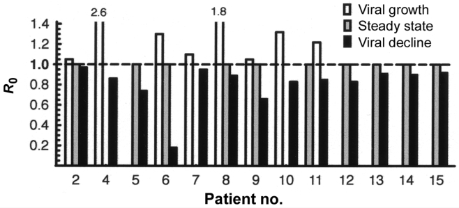
eration time of ∼50 h. For a lytically replicating virus such asBKV, if one assumes a fixed value of d between infection andthe bursting of progeny virions, the R is calculated by R p exp (sd) [11]. Applying a d of ∼48 h, we calculated R during viral growth (R 1 1) and decrease (R ! 1) after the respective interventions (figure 2). Three examples are discussed in moredetail. In patient 2, the R during the viral growth and decrease stages was 1.05 and 0.97, respectively. Therefore, switching fromTac and AZA to CsA and Aza around week 45 after transplan-tation had a 7% efficacy (R p 1.04–0.97). In patient 4, we Figure 2.
Efficacy of reduced immunosuppressive regimens. The basic observed a sharp increase in BKV plasma load after methyl- reproductive ratio (R ) is plotted during viral increase (white bars), steady prednisolone around day 80 (R p 2.6) that was followed by state (light gray bars), and viral decrease (dark gray bars). For patient 4, a decrease in BKV plasma load over the course of 60 days the R during viral growth was 2.6 ("rooftop value"). The dashed horizontal 0.86 , which indicated a 66% efficacy of switching from 1 indicates the critical threshold value separating viral growth from decrease.
Tac, Aza, and Pred plus methylprednisolone to low-dose Tacand leflunomide (LeF) but 14% efficacy relative to that ob- rate (t , 20 h), 1.09 ⫻ 10 –1.09 ⫻ 10 virions were produced served for the Tac, Aza, and Pred regimen before antirejection and cleared every day. To estimate the impact of this high in treatment with methylprednisolone. In patient 5, we observed vivo turnover, we assumed a BKV burst size of 1 ⫻ 103–1 ⫻ 104 a reduction in the R of 26% (from 1 to 0.74) after the change virions/lytically replicating host cell [12], which translates to in regimen around day 105 that did not persist; the R returned 1 ⫻ 10 –1 ⫻ 10 cells/day lysed through BKV replication alone.
quickly to ∼1, although a lower level of BKV replication was If this estimate of the number of infected cells is reasonably maintained. In patient 6, we observed a reduction in the R of 4 ⫻ 10 –4 ⫻ 10 BKV viruses are released from lysed 78% (from ∼1 to 0.22) around day 95. In patient 11, viral tubular epithelial cells every hour.
growth (R p 1.34) was observed after a steady state (R p Increases in BKV plasma load are of particular interest, be- 1 and treatment of a steroid-refractory interstitial rejection cause this may reflect the spread of BKV infection and the with coexisting PVAN with anti-lymphocyte globulin and a extent of tissue at risk. We found a median growth rate of 0.18/ switch from a Tac and Aza regimen to low-dose Tac and Sir.
day (t , ∼4 days), with the fastest being 0.90/day (t , 18 h) in Viral growth decreased (R p 0.85) after the switch to low- patient 4 and the slowest being 0.019/day (t , 37 days) in patient dose Sir and Aza. For patients 7–15, the respective efficacies 3 (table 2). In the densely sampled patient 1, a transient increase were as follows: patient 7, 13%; patient 8, 11%; patient 9, 34%; in BKV plasma load was noted in the first 4 h after nephrec- patient 10, 37%; patient 11, 30%; patient 12, 17%; patient 13, tomy, which was unlikely to have resulted from increased rep- 9%; patient 14, 10%; and patient 15, 8%. The mean (median) lication but may have been caused by surgical manipulations, efficacy for all patients and regimens was 27% (17%). Under as was described for Epstein-Barr virus load after resection of the assumption of an efficacy of 20% for reduced immuno- nasopharyngeal carcinoma [13].
suppressive regimens in a model patient with a quasi steady- On the basis of the rapid BKV clearance rate in vivo, cor- ) BKV plasma load of 1 ⫻ 10 copies/mL, a viral responding to a t of ∼2 h, and the intracellular delay d (the generation time of 2 days, and a detection limit of 500 copies/ viral eclipse phase between infection and the bursting of prog- mL, it would take ∼7 weeks for the BKV plasma load to decrease eny virions) in vitro of 48 h [14], we calculated a BKV gen- to below the limit of detection. For a BKV plasma load of Contrasting BK virus (BKV) kinetics with other known viral kinetics.
Baseline plasma viral load, copies/mL Clearance rate per daya Daily turnover,d % a Calculated by exponential decay slopes.
b Based on plasma viral DNA decay measurements.
c Based on plasma viral RNA decay measurements.
d Percentage of the total body virus population. References: simian immunodeficiency virus (SIV) [19]; HIV [17, 20]; hepatitis B virus (HBV) [21, 22]; hepatitis C virus (HCV) [20, 23, 24]; Epstein-Barr virus (EBV) [13]; cytomegalovirus (CMV) [25].
Polyomavirus BK Dynamics In Vivo • JID 2006:193 (1 January) • 85
1 ⫻ 10 copies/mL, it would take 13 weeks for the virus load plex combined antiviral interventions in future clinical stud- to decrease to below the limit of detection.
ies. If one assumes an average efficacy of 20% for an immu-nosuppressive regimen in a model patient with 1 ⫻ 10 or 1 ⫻ 107 BKV copies/mL at steady state, it would take ∼7 or 13 weeks,respectively, for the BKV plasma load to decrease to below the Mathematical models have contributed considerably to the un- limit of detection. These kinetics suggest that, for clinical man- derstanding of viral infections in vivo, including those with agement, biweekly monitoring in patients with reduced immu- hepatitis viruses, cytomegalovirus, and HIV-1 [15]. The com- nosuppression may be sufficient. However, we note that, after mon hallmark of these entities is progressive organ compromise reducing immunosuppressive regimens, there may be a delay of through persistent viral replication in the setting of immune 4–10 weeks in some patients before the BKV plasma load starts dysfunction. Although polyomavirus infections have not been to decrease (e.g., patients 13–15 in figure 1B).
studied so far, it is clear that the newly recognized PVAN shares The limitations of our study include the varying sampling these characteristics: it is a chronic progressive disorder accom- density, the relatively small sample size, and its retrospective panied by high BKV plasma loads in intensely immunosup- nature. However, the results were derived from careful analysis pressed renal allograft recipients. In clinical practice, PVAN is of multiple intervals of viral decay. The estimated kinetics of still viewed as a slowly progressing disease, although the mean BKV in PVAN lie in between the extremes of simian immu- time from diagnosis to allograft failure is only 11 months. Our nodeficiency virus [19] and HIV [17, 20], on the one side, and analysis indicated that rather rapid dynamics of BKV replication hepatitis B virus [21, 22], on the other (table 3), and are com- underlie the course of PVAN. Although effective BKV-specific parable to those of hepatitis C virus in chronic infection [20, antivirals are still lacking, the decrease in BKV plasma load af- 23, 24]. However, the smaller number of susceptible tubular ter allograft nephrectomy provided a unique opportunity to epithelial host cells, in addition to the complex situation present obtain the first minimal estimates of BKV clearance as being with allograft, may explain the faster progression to end-stage fast (t , 1–2 h) or moderately fast (t , 20–38 h). In accordance kidney failure in PVAN, compared with that in chronic hepatitis with the data, we assumed that the replication base for BKV C infection.
had been removed from the patient's body with the allograft In conclusion, we report the first evidence (to our knowl- nephrectomy. Of note, any residual replication would render edge) of fast and moderately fast replication kinetics of BKV these rates even faster. In patients with reduced immunosup- in renal transplant recipients. Our results emphasize organ cell pressive regimens, BKV clearance was moderately fast or slow, damage as a major pathogenetic factor in PVAN. Finally, the ranging from 6 h to 17 days. This variability is not fast BKV dynamics may explain the high frequency of BKV unexpected, given that different interventions were used and a genome rearrangements, which are unusual for DNA viruses number of complex factors may underlie this net decrease, in- and may be important for immune evasion, antiviral resistance, cluding the individual net state of immunosuppression and the and development of cancer.
quality and quantity of BKV-specific immune effectors [16].
On the basis of the study of renal biopsies by Randhawa et al. [12], who reported that BKV-infected renal cells release, on average, 6000 virions, and on the viral clearance rates that wecalculated, we estimated the daily tubular epithelial-cell loss We thank the patients and the transplant teams for participating in the resulting directly from BKV replication to be 1 ⫻ 10 –1 ⫻ 10 cells. However, the overall impact on allograft function is likelyto be underestimated, because this describes the average cyto- pathic aspect of PVAN without regarding the impact of focaltubular damage per nephron over the total of 1. Randhawa PS, Finkelstein S, Scantlebury V, et al. Human polyoma virus-associated interstitial nephritis in the allograft kidney. Transplan- nephrons and ignores other pathologic aspects of PVAN, such tation 1999; 67:103–9.
as immune-mediated damage.
2. Nickeleit V, Klimkait T, Binet IF, et al. Testing for polyomavirus type We propose the basic R as a measure to estimate the efficacy BK DNA in plasma to identify renal-allograft recipients with viral of complex interventions. The R is intimately related to viral nephropathy. N Engl J Med 2000; 342:1309–15.
3. Hirsch HH, Knowles W, Dickenmann M, et al. Prospective study of fitness in an individual transplant recipient [10, 15, 17]. The polyomavirus type BK replication and nephropathy in renal-transplant efficacies of reduced immunosuppression varied from 7% to recipients. N Engl J Med 2002; 347:488–96.
78% (mean, 28%; median, 22%), which are comparable to those 4. Ramos E, Drachenberg CB, Portocarrero M, et al. BK virus nephro- of cidofovir and LeF in vitro [18]. Because it is independent pathy diagnosis and treatment: experience at the University of Mary-
land Renal Transplant Program. Clin Transpl 2003; 143–53.
of the plasma viral load, R may prove to be an important and 5. Hirsch HH, Steiger J. Polyomavirus BK. Lancet Infect Dis 2003; 3:
much-needed in vivo measurement for the evaluation of com- 86 • JID 2006:193 (1 January) • Funk et al.
6. Fishman JA. BK virus nephropathy—polyomavirus adding insult to 16. Comoli P, Azzi A, Maccario R, et al. Polyomavirus BK-specific im- injury. N Engl J Med 2002; 347:527–30.
munity after kidney transplantation. Transplantation 2004; 78:1229–32.
7. Hirsch HH, Brennan DC, Drachenberg CB, et al. Polyomavirus- 17. Funk GA, Fischer M, Joos B, et al. Quantification of in vivo replicative associated nephropathy in renal transplantation: interdisciplinary anal- capacity of HIV-1 in different compartments of infected cells. J Acquir yses and recommendations. Transplantation 2005; 79:1277–86.
Immune Defic Syndr 2001; 26:397–404.
8. Drachenberg CB, Papadimitriou JC, Hirsch HH, et al. Histological 18. Farasati NA, Shapiro R, Vats A, Randhawa P. Effect of leflunomide and patterns of polyomavirus nephropathy: correlation with graft outcome cidofovir on replication of BK virus in an in vitro culture system. Trans- and viral load. Am J Transplant 2004; 4:2082–92.
plantation 2005; 79:116–8.
9. Hirsch HH. BK virus: opportunity makes a pathogen. Clin Infect Dis 19. Zhang L, Ribeiro RM, Mascola JR, et al. Effects of antibody on viral kinetics in simian/human immunodeficiency virus infection: impli- 10. Anderson RM, May RM. Infectious diseases of humans. New York: cations for vaccination. J Virol 2004; 78:5520–2.
20. Ramratnam B, Bonhoeffer S, Binley J, et al. Rapid production and Oxford University Press, 1991.
clearance of HIV-1 and hepatitis C virus assessed by large volume 11. Nowak MA, Lloyd AL, Vasquez GM, et al. Viral dynamics of primary plasma apheresis. Lancet 1999; 354:1782–5.
viremia and antiretroviral therapy in simian immunodeficiency virus 21. Nowak MA, Bonhoeffer S, Hill AM, Boehme R, Thomas HC, McDade infection. J Virol 1997; 71:7518–25.
H. Viral dynamics in hepatitis B virus infection. Proc Natl Acad Sci 12. Randhawa PS, Vats A, Zygmunt D, et al. Quantitation of viral DNA USA 1996; 93:4398–402.
in renal allograft tissue from patients with BK virus nephropathy. Trans- 22. Whalley SA, Murray JM, Brown D, et al. Kinetics of acute hepatitis B plantation 2002; 74:485–8.
virus infection in humans. J Exp Med 2001; 193:847–54.
13. To EW, Chan KC, Leung SF, et al. Rapid clearance of plasma Epstein- 23. Herrmann E, Neumann AU, Schmidt JM, Zeuzem S. Hepatitis C virus Barr virus DNA after surgical treatment of nasopharyngeal carcinoma.
kinetics. Antivir Ther 2000; 5:85–90.
Clin Cancer Res 2003; 9:3254–9.
24. Neumann AU, Lam NP, Dahari H, et al. Hepatitis C viral dynamics 14. Low J, Humes HD, Szczypka M, Imperiale M. BKV and SV40 infection in vivo and the antiviral efficacy of interferon-a therapy. Science 1998;
of human kidney tubular epithelial cells in vitro. Virology 2004; 323:
25. Emery VC, Cope AV, Bowen EF, Gor D, Griffiths PD. The dynamics 15. Perelson AS. Modelling viral and immune system dynamics. Nat Rev of human cytomegalovirus replication in vivo. J Exp Med 1999; 190:
Immunol 2002; 2:28–36.
Polyomavirus BK Dynamics In Vivo • JID 2006:193 (1 January) • 87

Source: http://www.herzzentrum-basel.ch/fileadmin/unispitalbaselch/Bereiche/Medizin/Infektiologie_Spitalhygiene/Paper_nicht_auf_PubMed/2007/6C2C4C80d01.pdf

Microsoft word - orthospec - faqs.doc

Orthospec™ ESWT Frequently Asked Questions (FAQs) Pre and Post Treatment Briefly describe the procedure (i.e., shockwave delivery, length of procedure, anesthesia, physician, technician, consumables, etc) The Orthospec™ utilizes electrohydraulic, spark gap technology. The treatment procedure is a non-invasive, outpatient procedure intended to treat chronic heel pain caused by Plantar Fasciitis. The treatment regimen calls for one 25 minute treatment session, providing a total of 3800 shocks. It is recommended that this procedure be performed by a qualified, Medispec trained, medical professional under the supervision of a physician. During the procedure, there is no anesthesia or sedation provided to the patient. Imaging is not required.