Layout
Onychomycosis and nail dystrophy treated with
the PinPointe FootLaserHamish Dow
A new modality has become available for
the treatment of onychomycosis. I wish to
share some results that I have achieved
using the new FootLaser made by PinPointe
the losing battle against this insidious
creates subungual damage, and a nutrient-
had some form of empirical testing and
disease, which is prevalent in the
rich source of decaying matter offers
reviews performed by the pharmaceutical
population worldwide. Pharmaceutical
avenues for pathogens to exploit.
industry. Prescription lacquers and oral
companies claim that as much as 12% of
Homeopathic and naturalistic remedies
remedies are example of OTC medication,
the population is contaminated with
such as bleach, tea tree oil, vinegar,
as are the range of sprays and creams
fungal nail disease.
thyme, oregano oil, ClariPro, Zetaclear,
overtly marketed as antifungal
The two cases I report both suggest
Antimonolium Curdum, Aloe Vera and
medications. The list includes terbinafine,
that tight-fitting shoes can cause
mouthwashes rarely work and are mostly
itraconizole, fluconazole, clotrimazole,
intermittent and prolonged trauma that
a waste of time and money. Pharmacology
tolnaftate, zinc undecenoate and
allows the skin defences to be breached.
is normally the most effective treatment
undecenoic acid.
Devitalisation of the local area through
for onychomycosis. Over-the-counter
The PinPointe FootLaser is the first
compressive exsanguination or tissue
(OTC) medication is available in outlets
medical device to obtain regulatoryclearance (US FDA, EU, Health Canada,Australia and others) for the safe andeffective treatment of onychomycosis. Thereported percentage of patients receivingbenefit from this treatment, 71.4%, issubstantial.1 Treatment with the FootLaseris repeatable, has no systemic toxicity andlaser treatment does not preclude the useof other modalities.2,3
In February 2009, I added the
FootLaser to my armamentarium ofremedies against this stubborn andpernicious disease. To date I have treatedover 150 patients. To follow are casereports of two of my patients, both withoutcomes that I have not seen before withother forms of therapy.
Independent analysis of data from 109
photos from 60 of my patients wasperformed in November 2010 anddemonstrated: that, at three months, 67%of all treated toes showed improvement,compared with 80% at six months, 68% atnine months and 84% at 12 months.
Again, the differences over time possibly
Figure 1. TOP. Patient 1 as she presented to my office in December 2009 with a clinical
reflect different patient populations as well
diagnosis and positive mycology of onychomycosis in all 10 toes. She received a single
as continued improvement.
treatment on 14 December and a second treatment in October 2010. BOTTOM. Appearance ofher nails at 13 months post-treatment. All 10 toes responded to treatment, most with 100%
The patient was an active and dynamic
PodiatryNow June 2011
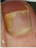
Figure 2. Time series composite of left (TOP) and right (BOTTOM) 1st toes of patient 1. The images show the rate and process of the lesiongrowing out over a period of 13 months.
smartly dressed, 66-year-old female withpronounced onychomycosis anddystrophyic nails on both feet; the right 1stnail (hallux) being the most severelyaffected. It was a condition she foundembarrassing, uncomfortable, emotionallydepressing and cosmetically abhorrent.
In around 2000, whilst out walking in
rough adventurous terrain, she damagedboth of her great toes to such an extentshe feared she would lose them. It wouldseem that a combination of poor toeboxspace and slack fastening of the footwearcaused repeated compression of the nail
Figure 3. Patient 1 right first toe before (left) and after (right) debridement.
plates, leading to subungual trauma andonycholysis.
Over the next two years she noticed
that they never fully recovered to their
nothing had worked to eradicate the
to keep her review appointments and that
former condition but began to ‘look
condition and was emotionally worn down
she needed to provide a high level of foot
worse'. The patient presented to a
by the effect of having a disfiguring
hygiene and antifungal care to prevent
podiatrist in 2008, where routine cutting
disease (as she saw it). A medical history
recolonisation by opportunistic pathogens.
and thinning of the nails was initiated
was documented, her feet were examined
The patient was also informed that
along with instructions on foot hygiene
and an explanation of the modality of the
recovery is highly variable from patient to
and advice to use OTC antifungal
PinPointe FootLaser was explained.
patient. Factors such as trauma, poor
preparations. Later in 2008, after lab
The patient was reminded of the
hygiene, poor circulation, duration and
confirmation of dermatophyte infection,
options available including
severity of the infection, age and general
the patient was prescribed oral terbinafine.
pharmacological, surgical and FootLaser. I
health may all influence recovery. Finally, I
As a consequence of the medication she
explained the benefit of a toxicity-free
told her that not every condition may
developed uncomfortable abdominal
approach, the fact that the FootLaser
resolve and a secondary treatment may be
swelling, giving her the appearance of
treatment is largely without any sensation,
required in about 15-20% of patients.
being some months pregnant and she
is condensed into the treatment time and
Following this explanation she consented
found it impossible to remain on the
has a level of success that surpasses all of
to having the treatment.
medication longer than two months.
the topicals and at least matches the
The patient's feet were cleaned and
By the time she arrived at my clinic in
outcome for oral medications, if not more
pre-treatment photographs were taken
December 2009 she was disheartened that
effective. I also explained the need for her
(Figures 1 & 2). All nails were debrided to
June 2011 PodiatryNow
traumatised the tissues. The right 1st toe
fitting stylish shoes is what is preventing
was displaying a transverse ridge with
the patient's recovery from reaching 100%,
dense keratinised tissue distally. All lesser
or at least preventing accurate visual
nails showed greater clarity within the
confirmation of 100% clearance.
structure and they cut with a crisp and
At this point, the evaluation of the nails
clear audible click, indicating improved
showed a change in texture vitality and
integrity. The left 1ST nail showed a more
structure. What was once a fungally
normal nail plate production in progress
infected, damaged and gryphotic right first
and an advancement of contamination
toenail has now returned to an almost
from the nail matrix of 2-3 mm.
normal nail in every way. The patient is
Importantly, the patient was very happy
enormously happy with her outcome. I am
with the early stages of the treatment. All
left with the satisfaction that the PinPointe
nails were cleaned and debrided to
FootLaser has safely and successfully
remove excess keratin and dystrophic
treated her fungal infection and in the
Figure 4. Continuous increase in percent
nail, and photographs taken. The patient
process has apparently stimulated a
clear nail over a 13-month period.
was instructed to continue with her
rejuvenation of the nail bed and tissues in
Photographs from each follow-up period
antifungal and hygiene care.
the nail root matrix. This is the first time I
were computer analysed to quantify the
The patient returned in July 2010 for
have seen this to occur in my entire career
change in area of clear nail. First left toe
her 7-month progress evaluation and nail
since graduating in 1981.
(BLUE) and 1st right toe (RED). The straight
care. Once more photographs were taken,
lines are linear regressions fit to the data
the nails reduced, onychophosis and
that indicate lesion clearing at a rate of
subungual debris removed and 1%
Patient 2 was a physically active working
about 6% of the total nail plate per month.
clotrimazole spray applied. Photographs of
male aged 63 with dystrophic and
the nails after the reductions were taken.
mycotically infected nails. The infection
By now the nails demonstrated significant
was clinically restricted to both 1st toes.
what I considered an optimal level. As
positive change and the most visually
Dermatophyte infection was confirmed by
much contaminated tissue needed to be
abnormal nail had almost grown out.
InTray DM at day 7 following initial
removed as possible and all debris cleared
In October 2010, 10-months post-
assessment, however his physician had
away, including subungual debris. This
treatment, the patient requested a second
also diagnosed it some time before. He
leaves a good working surface that offers
laser treatment ‘to be on the safe side,' as
was uncomfortable with the thickness of
less interference to the laser-energy
she put it. I advised that it was unlikely to
the nails and also dissatisfied by the
discharge and penetration into the tissues.
be necessary but the patient was far
physical appearance, particularly in
However, this is done in a fashion that
happier for me to apply the FootLaser
barefoot holiday settings. To the best of his
avoids trauma to the vital soft tissues.
again. This time only the margins and a
recollection he had had his condition for
Photographs were taken again after
single pass was performed over all nails.
‘several years'.
debridement (Figure 3).
Photographs were taken pre- and post-
During this time the patient had tried a
The FootLaser was methodically and
debridement antifungal spray applied.
wide variety of topical, proprietary nail
meticulously applied to all nails in a 1mm
In January 2011, 13 months after the
treatments and the condition continued to
spot matrix pattern. In addition to the nail
initial treatment, the patient once more
worsen. He did not consider oral
plate itself, the nail margins, nail-root
presented for routine care and an
medication as an option that he wished to
matrices and surrounding tissues were also
evaluation. Photographs were recorded
pursue due to its restrictions on
lased. The treatment area extended from
pre- and post-debridement and 1%
consumables and the impact that had on
approximately 4mm proximal from the
clotrimazole spray applied to protect the
his lifestyle. Having heard radio
eponychium to the end of the nail beds.
area. All nails show increased clarity,
advertisements about the new FootLaser,
The pattern was repeated twice over each
integrity and health (Figure 1).
he arrived for treatment at my clinic on 17
November 2009.
Following the laser treatment,
Analysis and discussion
His pastimes included competitive
terbinafine spray was applied to the nails,
I sent my before and after photographs of
squash, extensive hill and robust fell
toes and interdigital areas and terbinafine
the patient's left and right 1st toes to a
walking. An examination of his footwear
cream was applied to the skin on the
research company that uses a trained
indicated the need for a larger size but
plantar surfaces to help prevent
technician and a computer algorithm to
complications were created by the
recolonisation. The post-care advice was
measure the area of clear nail on each
narrowness of his feet causing difficulties
explained, and an advice sheet given
image. The results are presented in
in obtaining the best fit, as length is
together with antifungal cream and spray.
Figure 4. What is plotted is the percent of
sometimes sacrificed for a narrower width
A letter was sent to her GP detailing the
clear nail measured from each follow-up
to ensure better grip on the foot. I
treatment and contact made with her
photograph before debridement. The left
explained the need for improved toebox
current podiatrist explaining the
toe (blue) appears stalled at the onset,
space during the post-laser recovery
then, at 7-months it ‘catches up' with the
In March 2010 the patient returned for
right. Both nail plates grow clear nail at a
On examination there was gross
a three-month interim follow-up
rate of about 6% of the nail plate surface
thickening of both 1st toe nail plates
examination. At this point and it was
per month. This is about the same growth
consistent with prolonged trauma and
apparent that there were positive
rate as the nail plate itself.
substantial subungual debris. This material
improvements in the nails, although
I also noticed from this analysis that
was harvested for culture, which
bruising was evident in the nail due to the
the clearing seems to stop at about 80%.
patient choosing stylish footwear that
Perhaps the continued trauma from tight-
contamination by day 7.
PodiatryNow June 2011
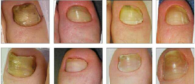
Figure 5. Time series composite of left (TOP) and right (BOTTOM) 1st toes of patient 1. The images show the rate and process of the lesiongrowing out over a period of 13 months.
A medical history was documented and
structural integrity and improved clarity
this is certainly not the case since the
an explanation of the modality of the
within the structure. When cut, all nails
patient is extremely pleased with the
PinPointe FootLaser explained. Then the
yielded a crisp and clear audible click and
overall cosmetic improvement. He has had
consent forms were completed and pre-
had good tensile strength, indicating
a lengthy period of time (July 2010 until
treatment photographs taken (Figure 5).
improved integrity. Both 1st nails showed
March 2011) without any clinical
All nails were extensively debrided and all
an improved connection to the nail bed at
interventions of any type and his nails
loose subungual debris removed using
the distal margin. The nail on the first toe
have continued to show improvement in
clippers and Podospray foot drill with
on the left foot showed a visible band of
texture, clarity and health. The only
combinations of tungsten carbide burrs.
3mm width, indicating a difference in its
negative I can find is the damage done to
The FootLaser was methodically and
structure compared with the rest of the
his nails from impact from his physical
meticulously applied to all nails in a
nail. I speculated that this was perhaps
activities. I have yet to persuade him that
lateral then longitudinal 1mm spot matrix
newly re-keratinising nail plate and nail
his feet are longer than he thinks.
pattern. The nail margins, nail-root
I am impressed with the changes that
matrices and surrounding tissues were
All nails were cleaned and debrided to
occurred in the nails of this patient
lased proximal. Treatment began at
remove excess keratin and dystrophic nail
following one intervention with the
approximately 4mm from the eponychium
and photographs taken. The patient was
PinPointe FootLaser. It has led me to
and extended to the very end of the nail
instructed to continue with his antifungal
believe that the laser has some other (as
beds. All 10 toes were treated and
hygiene care. The patient was comfortable
yet unknown) rejuvenating influence on
particular attention was applied to both 1st
and satisfied with the look at this early
the germinating tissues. Although I have
nails. The patient expressed his relief at
no direct evidence, my own personal
the procedure being entirely painless.
The patient returned in July 2010, 8-
experience with the laser and my years as
Following the laser, terbinafine spray
months post-treatment for his second
a practising clinician lead me to believe as
was applied to the nails, toes and
progress evaluation, and nail care. Once
I continue to work with the laser that I am
interdigital areas, and then terbinafine
more photographs were taken, the nails
witnessing treatment outcomes that have
cream was applied to the skin on the
reduced and onychophosis and subungual
not been witnessed before.
plantar surfaces. The post-care advice was
debris removed and 1% clotrimazole spray
explained, and an advice sheet given along
applied. Photographs of the nails after the
with antifungal cream and spray. A letter
reductions were taken.
1. Uro M, L Uro, M Abrahams, M Abrahams, R
was sent to his GP detailing the treatment
By now the nails had already
Grzywacz. Safety and efficacy of FootLaser
and contact made with his current
demonstrated significant positive change
treatment of onychomycosis in private
podiatrist explaining the procedure.
and most visually abnormal nail had
practice. Lasers Surg Med 2011; 43(S23):
In March 2010 the patient returned for
almost completely grown out (Figure 5).
954 (abstr).
2. Abrahams M, Dow H, Grzywacz R, Uro M,
a 4-month post-treatment interim
On February 11 2011, 15 months
Harris DM. Efficacy of FootLaser treatment
examination. At this point new
following FootLaser treatment the lesions
of onychomycosis in 262 private practice
photographs were taken, and it was
in most nails had completely grown out.
patients. 2011. Pending submission.
apparent that there were positive
Yet, the computer analysis on this patient
3. Harris DM, J Strisower, B McDowell. Pulsed
improvements in the nails. Although the
returned ‘100% infected' due to the
laser treatment for toenail fungus. SPIE
nails were still thick they showed good
persistent whitish discoloration. However,
Proceedings 7161A-121, 2009.
June 2011 PodiatryNow
Source: http://questlight.net/wp-content/uploads/2016/02/Onycomycosis-White-paper.pdf
Osteoporos IntDOI 10.1007/s00198-011-1528-y Skeletal mineralization defects in adulthypophosphatasia—a clinical and histological analysis F. Barvencik & F. Timo Beil & M. Gebauer & B. Busse &T. Koehne & S. Seitz & J. Zustin & P. Pogoda & T. Schinke &M. Amling Received: 14 April 2010 / Accepted: 3 January 2011 # International Osteoporosis Foundation and National Osteoporosis Foundation 2011
Deaths from Marijuana v. 17 FDA-Approved Drugs (Jan. 1, 1997 to June 30, 2005) http://medicalmarijuana.procon.org/view.resource.php?resourceID=000145 I. Background Much of the medical marijuana discussion has focused on the safety of marijuana compared to the safety of FDA-approved drugs. On June 24, 2005 ProCon.orto the US (FDA) to find the number of deaths caused by marijuana compared to the number of deaths caused by 17 FDA-approved drugs. Twelve of these FDA-approved drugs were chosen because they are commonly prescribed in place of medical marijuana, while the remaining five FDA-approved drugs were randomly selected because they are widely used and recognized by the general public.