Jdd 7-3 cover 1-4:cover
COPYRIGHT 2008 JOURNAL OF DRUGS IN DERMATOLOGY
INCREASE IN COLLAGEN TURNOVER INDUCED BY
INTRADERMAL INJECTION OF CARBON DIOXIDE IN RATS
Julio Cesar Tavares Ferreira MD,a Alessandra Haddad MD PhD,b Simone Arruda Navarro Tavaresc
a. General Surgeon, Member of the Brazilian College of Surgeons, Member of Brazilian Medical Society for Intradermal Therapy
b. Plastic Surgeon, MSc in Reconstructive Plastic Surgery from the Federal University of São Paulo – Escola Paulista de Medicina,
Member of the Brazilian Society of Plastic Surgeons
c. Physiotherapist, Specialist in Dermatologic-Functional Physiotherapy from Faculdade Integrada do Ceará;
Member of the Brazilian Aesthetics Academy
Abstract
Introduction: Results from clinical observations have demonstrated that percutaneous infiltration of carbon dioxide im-
proves the appearance of the skin in adjacent areas. No studies have been found in the literature that showed evidence
of histological changes caused by carbon dioxide injections.
Objectives and Methods: A blind cross-sectional pilot study was performed in the Departments of Pharmacology and
Morphology of the Federal University of Ceará, with the aim of histologically investigating whether intradermal and/or
subcutaneous injection of medicinal carbon dioxide would increase collagen turnover in rats. Ten male Wistar rats were
used, aged 3 months (2 animals) and 14 months (8 animals). The 2 younger rats were used as controls. Four of the older
rats received injections of saline solution (0.9%), and were also considered to be controls. In the remaining 4, carbon
dioxide was injected into the subcutaneous cellular tissue and intradermally. Biopsy samples were collected before and
after treatment with carbon dioxide.
Results: Collagen turnover increased in the treated animals in comparison with the controls. Compression of collagen
bundles in the tissue samples where intradermal injection was used was more intense than in the subcutaneous treatment.
The histological characteristics of the samples with carbon dioxide injected intradermally were similar to the characteristics
of the younger rats (controls).
Conclusions: The results obtained corroborate clinical observations of aesthetic improvements in the facial skin with car-
bon dioxide injections. Future research should address the comparison between intradermal and subcutaneous injections,
the volume of gas used, and the frequency of treatment sessions.
medium through which nutrients are offered to cells and into
Human tissue changes with age. In the skin, these modifi-
which cellular excreta are ejected.21,22 The extracellular ma-
cations are more easily recognized.1,2 The modifications to the
trix is composed of fluid and fibrous components. One of the
collagen and elastin system cause wrinkles, atrophy, grooves,
most common fluid components of the ECM is hyaluronic
ptosis, and laxity. These are the most evident signs of old
acid. In combination with certain proteins, this becomes a
skin,3-6 which can result in significant psychological problems
highly viscous and hydrophilic mucopolysaccharide. Elastin,
for individuals. There is increasing interest in therapeutic
fibronectin, and collagen are the basic elements of the fibrous
resources for solving such problems.7 For this reason, the mor-
component of the dermis. Collagen is the most abundant of
phological and structural changes to the dermis related to
these, and it is the main element of human skin. It is re-
aging are constantly being studied.
sponsible for maintaining the structural integrity of the skinby joining cells to other cells and the ECM.
Many authors have shown that there are beneficial effectsfrom subcutaneous carbon dioxide (CO
Collagen is an insoluble protein that forms fibers, which in
2) therapy for several
clinical conditions.7-20 Intradermal injection of CO
turn join together in bundles. Fibroblasts secrete the pre-
been studied recently, especially because of the clinically fa-
cursors of collagen, protocollagen types 1 and 3. Aging brings
vorable results observed, its low cost, and safety.8 However,
a decrease in the number of skin fibroblasts and, at the same
there is a lack of scientific evidence regarding the histolog-
time, an increase in collagenases, which catalyze the degra-
ical modifications that occur with the intradermal injection
dation of collagen. It also causes a reduction in the number
of cells and vessels and this, together with biochemical
changes in collagen constitution, leads to thinner collagen
fibers. The collagen content per unit of skin area decreases
Senile dermal modifications occur mainly in the extracellu-
by 1% per year over the course of adult life and collagen fibers
lar matrix (ECM), a substance within the extracellular space
become disorganized, less compact and more granular; col-
that provides the supporting structure to cells, as well as
lagen becomes more rigid and less elastic. The consequence
resistance to compression and stretching. It is also the
of this is the loss of dermal volume in older individuals.
JOURNAL OF DRUGS IN DERMATOLOGY
INCREASE IN COLLAGEN TURNOVER INDUCED BY
MARCH 2008 • VOLUME 7 • ISSUE 3
INTRADERMAL INJECTION OF CARBON DIOXIDE IN RATS
Some other clinical manifestations of aging, such as wrinkles,
contraindicated for phlebitis, cardiac/respiratory insuffi-
grooves, and atrophy, as well as changes in facial format, are
ciency, renal/hepatic insufficiency, severe arterial hyperten-
due to the new architecture of the skin's conjunctive tissue.23-35
sion, and pregnancy.8,10
Aging is a progressive, universal process that is subject to en-vironmental, genetic, and hormonal factors.7 Among the
Carbon dioxide provokes local vasodilation and increased re-
environmental factors associated with skin aging, ultraviolet
gional blood flow and oxygen pressure; there is a reduction in
radiation is the most important.36,37
the affinity of hemoglobin for oxygen, thereby resulting in moreavailability of oxygen for the tissue.36 Many authors have ob-
served this phenomenon after subcutaneous injection of CO2,
Many treatments have been used to reduce the effects of the
using many different examination techniques.12,15-17,40-42
skin aging process. Antiaging treatments have included
tretinoin (retinoic acid), antioxidants like vitamins C and E,phytoestrogens, dimethylaminoethanol, and many others in
This study was designed to investigate whether CO2 injec-
the form of creams, gels, and lotions. They can be used pro-
tion into the dermis of Wistar rats can improve collagen
phylactically, as coadjuvant therapies in some circumstances,or as the sole treatment. Invasive techniques are also availableand these seem to be more efficient, including chemical or
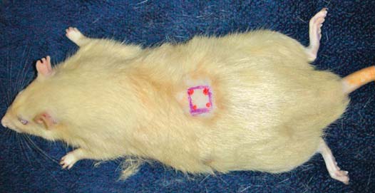
Figure 1a. Procedures undertaken with the 2 younger Wistar rats (3
laser peeling, dermal filling with various substances or with
months old, white circles) and the 8 older rats (14 months old,black/gray circles): GTI (intradermal CO infiltration), GTS (sub-
solid implants, botulinum toxin, and many plastic surgery
cutaneous CO infiltration), or infiltration of saline solution.
techniques. More recently, hormonal modulation has been
proposed as a prophylactic method,38 and CO2 therapy is also
being used as a promising technique against skin aging.8
Carbon Dioxide Therapy
Subcutaneous administration of CO2 is popularly known as
carboxitherapy.8,10 Subcutaneous infiltration is a recent in-novation in medicine, but the administration of CO2 began
in France in the 1930s, where peripheral arteriopathy wastreated with CO2 gas.10,12,39,40 The publication of studies on
CO2 therapy began in the 1950s, but most of the work papers
were published between 1985 and 2002.12,14
The main indications for CO2 therapy are peripheral arteri-
opathy,14,39 acrocyanotic syndrome,19 venous insufficiencyand foot and leg ulcers,13,41 adipose tissue accumulation,18,42symmetrical multiple lipomatosis,18 and others.
In the 1990s, video laparoscopy was used for injecting quan-tities of more than 3 liters of CO2,8,43 at rates reaching 1 liter
per minute without adverse effects.43 Thus, the use of this gas,which is formed naturally by the body at rest and during ex-ercise,10,42 is safe. Moreover, it is eliminated within a shorttime. Adjacent to the areas where CO2 was injected, surgeons
noticed reductions in the quantities of adipose tissue and fibrous edema.8,15 This prompted new clinical research usingsubcutaneous CO2 to treat adiposity and aging.7,9,18
A histological study on patients treated with CO
Figure 1b. Example of infiltration and punch biopsy site.
tions for adiposity showed that the treatment did not lead toany damage affecting the connective tissue, vascular bed, ornerve structures.15 The recent use of CO2 in angiographic
procedures has proven the safety of this cheap and nonal-lergenic gas.44 Carbon dioxide does not provoke embolismeven with bolus injections of 100 ml, and a continuous fluxof 20 to 30 ml/second does not induce adverse reactions.45Local or systemic complications have not been reported inthe literature.11,15,46 The only side effects are pain, hematomascaused by the puncture, and a crepitation sensation causedby the small local subcutaneous emphysema, which disap-pears within 30 minutes.7,8 Carbon dioxide injections are
JOURNAL OF DRUGS IN DERMATOLOGY
INCREASE IN COLLAGEN TURNOVER INDUCED BY
MARCH 2008 • VOLUME 7 • ISSUE 3
INTRADERMAL INJECTION OF CARBON DIOXIDE IN RATS
turnover and compare injections in the subcutaneous and
Biopsy and Histological Analysis
intradermal layers.
On the sixth day after the CO2 infiltration therapy, the 8
14-month-old rats (4 treated and 4 controls) were again
anesthetized with ether and the total skin thickness was
This pilot study was a blind, interventional, cross-sectional
biopsied using a 1-mm punch at the center of the 1-cm
study, with qualitative analysis of the results, carried out in
squares. Both the preprocedure biopsy specimens (from all 10
the Pharmacology and Morphology Departments of the Fed-
animals) and the biopsy specimens collected after the pro-
eral University of Ceará (UFC), from February to March in
cedure (from the treatment group) were prepared for histo-
2006. The experimental study followed the principles for
logical analysis.
research using animals: the subjects had full access to waterand food before and after the procedure and their life cycles
The specimens were fixed in formaldehyde and dehydrated in
(diurnal and nocturnal) were respected. The study was ap-
a series of increasing concentrations of ethyl alcohol. After
proved by the Ethics Committee for Animal Research of this
this, they were diaphanized, embedded in paraffin blocks and
university. The animals were provided by the Pharmacology
cut into 4-mm sections. The specimens were then subjected
Department of UFC.
to Mallory's trichrome staining, in which collagen synthesis
Ten male Wistar rats, born from the same mother and father,2 of them young (3 months old) and 8 of them old (14
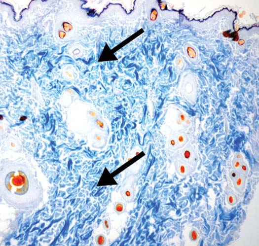
Figure 2. Photomicrograph of Wistar rat dermis biopsy a) before
months old), were used. The rats were subjected to a skin
and b) after subcutaneous CO infiltration, with less dispersed and
more numerous collagen fibers.
biopsy 1 day before the procedure and those presenting withdermatological or subcutaneous problems were excluded.
After either subcutaneous or intradermal CO
the morphological alterations (quantity and arrangement) tothe dermal collagen fibers were evaluated (Figure 1a).
Preprocedure Biopsy
After undergoing anesthesia (by means of inhaling ether
from a cotton ball inside a ventilated chamber), all 10 ani-
mals (young and old) were shaved and skin samples were
collected by punch biopsy (1 mm diameter in the right
posterior flank, 3 cm distally from the femoral joint).
Procedure
One day after the first biopsy, areas of 1 cm2 were shaved and
marked out on the skin of each of the 8 14-month-old rats
(Group T=treated): 1 area on the left anterior flank and the
other on the right anterior flank (3 cm posteriorly to the
scapulohumeral joint).
A disposable needle (30 G 1/2) was attached to the Carbtekcarboxitherapy equipment (Estek, São Paulo, Brazil) bymeans of an appropriate device with an anti-reflux filter. Themanometer pressure was calibrated to 15 mmHg, with a flowvelocity of 20 mmHg.
For 4 of the 14-month-old rats, CO2 infiltration was performed
intradermally (GTI) on the right side. This was done by meansof introducing only the bevel of the needle into 1 of the ver-tices of the square that was marked out, until the intradermalinfiltration of CO2 caused a white papule in the center of the
marked square. On the left side of the same rats, the injectionwas subcutaneous (GTS), with 3 mm of the needle introducedfrom 1 of the vertices of the square marked out, until the gasvolume was enough to cause skin distension in the marked area.
The point of the needle was kept in the corner of the square topreserve the central area for puncture biopsy (Figure 1b).
For the other 4 14-month-old rats (control group), injectionof saline solution (0.9%) into the subcutaneous tissue causeddistension of the areas marked out with squares.

JOURNAL OF DRUGS IN DERMATOLOGY
INCREASE IN COLLAGEN TURNOVER INDUCED BY
MARCH 2008 • VOLUME 7 • ISSUE 3
INTRADERMAL INJECTION OF CARBON DIOXIDE IN RATS
is shown in blue. The analysis was performed by an experi-
identification of each animal specimen (ie, whether it was
enced pathologist using a binocular optical microscope with
from the treated or the control group).
millimeter scale (Leitz Periplan GF 100X, Wetzlar, Germany).
Digital photos of the histological slices were taken (Collpix
camera, Nikon, Japan). The pathologist was not aware of the
No side effects were observed after the injection of CO2 or
saline solution.
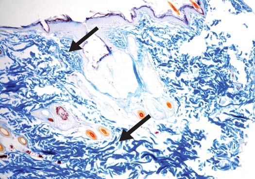
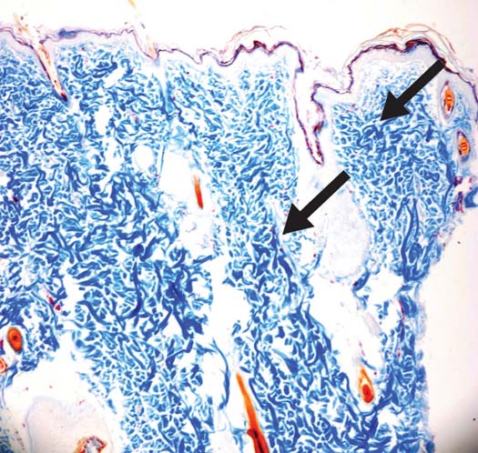
Figure 3. Photomicrograph of rat dermis biopsy after intradermal
Histological analysis showed intense collagen turnover in
CO infiltration in a) 14-month-old Wistar rat and b) 3-month-old
the skin samples of the animals treated with CO2, especially
in those with intradermal treatment, in comparison with theanimals that had only had saline solution injected in the der-
mis. Collagen synthesis was shown by Mallory's staining. Thecollagen fibers were less dispersed (Figure 2) following bothsubcutaneous and intradermal CO2 infiltration. Intradermal
injection made the collagen fibers more compact than did subcutaneous injection. After intradermal infiltration, the col-lagen arrangement in the dermis of the old animals in thisstudy was similar to that of the 2 young rats (Figure 3).
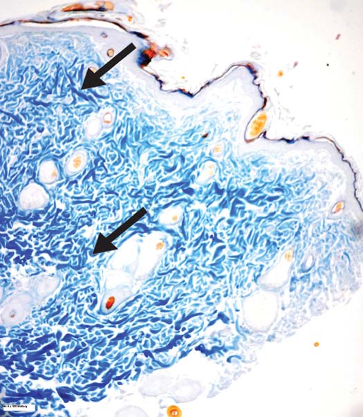
Subcutaneous saline solution injection into the control grouprats caused greater collagen fragmentation, comparing thepreprocedure biopsy with the biopsy after the procedure inthe control group (Figure 4).
Discussion
This study showed visible compacting of collagen fibers fol-
lowing CO2 infiltration, especially among the skin samples
with intradermal infiltration. Oria et al (2003), in a studyusing cadavers, had already demonstrated that aging causedthe collagen bundles to disperse, since collagen fibers aremore compact in young humans.47 In fact, after CO2 injection,
the arrangement of the collagen in the dermis of the older ratsin this study was similar to that of the younger animals.
Figure 4. Photomicrograph of rat dermis biopsy after saline solu-
tion infiltration in a 14-month-old Wistar rat (control).
JOURNAL OF DRUGS IN DERMATOLOGY
INCREASE IN COLLAGEN TURNOVER INDUCED BY
MARCH 2008 • VOLUME 7 • ISSUE 3
INTRADERMAL INJECTION OF CARBON DIOXIDE IN RATS
Many authors have shown that injection of CO
3. Lavker RM, Zheng PS, Dong G. Morphology of aged skin. Clin
subcutaneous layer leads to an increase in the blood flow to
Geriatr Med. 1989;5:53-67.
the area.12,15-17,40-42 This blood flow increase may lead to
4. Tsuji T, Yorifuji T, Hayashi Y, Hamada T. Light and scanning elec-
neoangiogenesis in cases of chronic exposure to CO
tron microscopic studies on wrinkles in aged persons' skin. Br J
tion,14 and could partially explain the more intense collagen
synthesis adjacent to the treated area in this study. In a ran-
5. Castelo-Branco C, Pons F, Gratacos E, et al. Relationship between
domized, controlled study by Brandi et al, subcutaneous
skin collagen and bone changes during aging. Maturitas. 1994;18:
injections of CO
2 to treat adiposity caused an increase in oxy-
gen pressure, and skin thickness, and a decrease in the cir-
6. Vitellaro-Zuccarello L, Cappelletti S, Dal Pozzo Rossi V, Sari-Gorla
cumference of the treated areas. A histological examination
M. Stereological analysis of collagen and elastic fibers in the nor-
showed that the adipocyte membrane was broken, and that
mal human dermis: variability with age, sex, and body region. Anat
connective tissue, vessels, and nerves were preserved.15 Other
studies are necessary to confirm these findings and the exact
7. Kede MPV, Sabatovich O. Dermatologia Estética. Rio de Janeiro:
mechanism of action. It may be necessary to use hyaluronic
Atheneu; 2004.
acid together with CO2 therapy to enhance results.
8. Lopez JC. Carbon Dioxide Therapy. University Hospital of Siena:
Italy; 2005.
Elastic fibers break during the process of aging,47 thereby caus-
9. de Souza RA, Garcez CE. Temas de Medicina Estética. 5ed. Porto
ing harm to dermal tissue and flaccidity. Recent clinical ob-
Alegre: Artes Médicas; 2005.
servations have shown that CO2 therapy can be used to
10. Brockow T, Hausner T, Dillner A, Resch KL. Clinical evidence of
treat laxity8 and that the intradermal layer is the best place
to perform gas injection, as demonstrated in this study. It re-
2 insufflations: a systematic review. J Altern Com-
plement Med. 2000;6:391-403.
mains to be seen whether this subtle but important change
11. Savin E, Bailliart O, Bonnin P, et al. Vasomotor effects of tran-
in the place of injection is capable of inducing elastic fiber
production (Figures 2 and 3). The present study did not aim
2 in stage II peripheral occlusive arterial disease.
to evaluate the quantity of collagen fibers nor the volume of
12. Ito T, Moore JI, Koss MC. Topical application of CO
gas to be injected, but simply to investigate whether CO
skin blood flow. J Invest Dermatol. 1989;93:259-262.
injection could increase collagen turnover. Other work in thescientific literature with this objective was found. Brandi et
13. Grosshans A, Gensch H. CO2-Gasinjektion--Indikation und
al (2001)15 showed that there was an evident increase in the
2 gas injection--indications and results]. Z Gesamte
Inn Med. 1987;42:667-670.
dermal thickness of patients treated for adipose accumula-tions using subcutaneous CO
14. Ambrosi C, Delanoe G. Action thérapeutique du CO2 naturel in-
2 injections. Lipolysis may, in
jecté sous la peau dans les artériopathies des membres. Etude ex-
such cases, be associated with increased collagen turnover.
périmentale. [Therapeutic effect of CO2 injected sub-cutaneously
Randomized trials may help to increase knowledge and
in arteriopathies of the limbs. Experimental research (author'stransl)]. Ann Cardiol Angeiol (Paris). 1976;25:93-98.
establish scientific parameters for the use of CO2 therapy.
Future research should use a larger number of animals (pos-
15. Brandi C, D'Aniello C, Grimaldi L, et al. Carbon dioxide therapy
sibly rabbits, whose skin is more similar to human skin) for
in the treatment of localized adiposities: clinical study andhistopathological correlations. Aesthetic Plast Surg. 2001;25:170-174.
studying CO2 therapy for aesthetic problems relating toaging skin, and try to standardize. The frequency of CO
16. Colin C, Lagneaux D, Lecomte J. Effets vasodilatateurs du CO2
injection sessions and the number of sessions needed, the
sec au niveau des vaisseaux cutanés. [Local vasodilatating effects ofcarbon dioxide on cutaneous blood vessels (author's transl)]. J Belge
spacing between the injection points on the skin, and the
Med Phys Rehabil. 1978;1:326-334.
volume of gas to be injected.
17. Bartoletti CA, Parassoni L, Varlaro V. La carbossiterapia: una
metodica terapeutica in evoluzione. Rivista La Medicina Estetica.
1997;2. Available at: http://www.lamedicinaestetica.it/Medest/
The data from this pilot study suggested that CO2 injection
RisultatoAbstract.asp. Accessed on March 29, 2007.
caused increased collagen turnover in rats, which was more
18. Brandi C, Bacci PA, Lattarulo P, et al. "Il trattamento chirurgico
pronounced with intradermal injection. These results support
delle localizzazioni addominali della lipomatosi multlipa simmet-
the clinical observation of reductions in wrinkles consequent
rica (LSM) integrato dalla carbossiterapia"; Unità Operativa di
to CO2 therapy and also establish that intradermal injection
Chirurgia Plastica e Ricostruttiva, Università degli Studi di Siena;
is better than subcutaneous injection.
48° Congresso Nazionale della Società di Chirurgia Plastica ed Es-tetica, Gubbio, 25-30 settembre 1999.
19. Toriyama T, Kumada Y, Matsubara T, et al. Effect of artificial car-
1. Gonçalves AP. Envelhecimento cutâneo cronológico. [The cuta-
bon dioxide foot bathing on critical limb ischemia (Fontaine IV)
neous chronologic aging (CCA)]. An Bras Dermatol. 1991;66:4s-6s.
in peripheral arterial disease patients. Int Angiol. 2002;21:367-373.
2. Trelles MA, Rigau J, Mellor TK, Garcia L. A clinical and histo-
20. Zwaan M, Kloess W, Kagel C, et al. Kohlendioxid als alternatives
logical comparison of flashscanning versus pulsed technology in
Kontrastmittel für die periphere Angiographie. [Carbon dioxide
carbon dioxide laser facial skin resurfacing. Dermatol Surg. 1998;
as an alternative contrast medium in peripheral angiography]. Rofo.
JOURNAL OF DRUGS IN DERMATOLOGY
INCREASE IN COLLAGEN TURNOVER INDUCED BY
MARCH 2008 • VOLUME 7 • ISSUE 3
INTRADERMAL INJECTION OF CARBON DIOXIDE IN RATS
21. Douglas CR. Tratado de fisiologia — Aplicado às ciências médicas. Rio
41. Schnizer W, Erdl R, Schops P, Seichert N. The effects of external
de Janeiro: Guanabara Koogan; 2007.
CO2 application on human skin microcirculation investigated by
22. Guyton AC. Tratado de fisiologia médica. 10th ed. Rio de Janeiro:
laser Doppler flowmetry. Int J Microcirc Clin Exp. 1985;4:343-350.
Guanabara Koogan; 2002.
42. Albergati F, Currì SB, Guidi F, et al. Effetti sul Microcircolo di dif-
23. Leveque JL, Corcuff P, de Rigal J, Agache P. In vivo studies of the
ferenti dosi di CO2 nella pannicolopatia edemato-fibrosclerotica
evolution of physical properties of the human skin with age. Int J
da stasi ("Cellulite"). Rivista La Medicina Estetica. 1998;2. Available
at: http://www.lamedicinaestetica.it/Medest/RisultatoAbstract.asp.
Accessed on March 29, 2007.
24. Macedo OR. A construção da beleza. São Paulo: Globo; 2005.
43. Vilos GA, Vilos AG. Safe laparoscopic entry guided by Veress nee-
25. Junqueira LCU, Carneiro J. Histologia básica. 8th ed. Rio de
dle CO2 insufflation pressure. J Am Assoc Gynecol Laparosc. 2003;
Janeiro: Guanabara Koogan; 1995.
26. Prockop DJ, Kivirikko KI, Tuderman L, Guzman NA. The biosyn-
44. Shaw DR, Kessel DO. The current status of the use of carbon diox-
thesis of collagen and its disorders (first of two parts). N Engl J
ide in diagnostic and interventional angiographic procedures. Car-
diovasc Intervent Radiol. 2006;29:323-331.
27. Cruzzi-Maya T. Pineiro-Maceira J. Dermatopatologia: bases para o
45. Lang EV, Gossler AA, Fick LJ, et al. Carbon dioxide angiography:
diagnóstico morfológico. São Paulo: Roca; 2001.
effect of injection parameters on bolus configuration. J Vasc Interv
28. Tsuji T, Hamada T. Age-related changes in human dermal elastic
fibres. Br J Dermatol. 1981;105:57-63.
46. Ochiai R, Takeda J, Noguchi J, et al. Subcutaneous carbon diox-
29. Blair C. Morphology and thickness of the human stratum
ide insufflation does not cause hypercarbia during endoscopic thy-
corneum. Br J Dermatol. 1968;80:430-436.
roidectomy. Anesth Analg. 2000;90:760-762.
30. Lapiere CM. The ageing dermis: the main cause for the appear-
47. Oriá RB, Brito GAC, Ferreira FVA, et al. Estudo das alterações
ance of ‘old' skin. Br J Dermatol. 1990;122(suppl 35):5-11.
relacionadas com a idade na pele humana, utilizando métodos dehisto-morfometria e autofluorescência. [Study of age-related
31. Kono T, Tanii T, Furukawa M, et al. Correlation between ageing
changes in human skin using histomorphometric and autofluores-
and collagen gel contractility of human fibroblasts. Acta Derm
cence approaches]. An Bras Dermatol. 2003;78:425-434.
32. Moragas A, Garcia-Bonafe M, Sans M, et al. Image analysis of der-
mal collagen changes during skin aging. Anal Quant Cytol Histol.
ADDRESS FOR CORRESPONDENCE
33. Ashcroft GS, Greenwell-Wild T, Horan MA, et al. Topical estro-
Julio César Tavares Ferreira
gen accelerates cutaneous wound healing in aged humans associ-
ated with an altered inflammatory response. Am J Pathol. 1999;155:
1505/1302 CEP 60-125-150
Fortaleza - Ceará
34. Takeda K, Gosiewska A, Peterkofsky B. Similar, but not identical,
modulation of expression of extracellular matrix components dur-
ing in vitro and in vivo aging of human skin fibroblasts. J Cell Phys-iol. 1992;153:450-459.
35. Pienta KJ, Getzenberg RH, Coffey DS. Characterization of nuclear
morphology and nuclear matrices in ageing human fibroblasts.
Mech Ageing Dev. 1992;62:13-24.
36. Bernstein EF, Chen YQ, Kopp JB, et al. Long-term sun exposure al-
ters the collagen of the papillary dermis. Comparison of sun-protectedand photoaged skin by northern analysis, immunohistochemical stain-ing, and confocal laser scanning microscopy. J Am Acad Dermatol.
1996;34:209-218.
37. Margelin D, Fourtanier A, Thevenin T, et al. Alterations of pro-
teoglycans in ultraviolet-irradiated skin. Photochem Photobiol.
1993;58:211-218.
38. Life JS, Mintz AP. Menopause and andropause. Marking the rest of
our life the best of our life. Available at: http://agemanagement-center.com/pdf/menapause_and_andropause.pdf. Accessed onMarch 29, 2007.
39. Fabry R, Dubost JJ, Schmidt J, et al. Le traitement thermal des
maladies artérielles: un placebo coûteux ou une thérapeutique àpart entière? [Thermal treatment in arterial diseases: an expensiveplacebo or an effective therapy?]. Therapie. 1995;50:113-122.
40. Belotti E, Bernardi M. Utilizzazione della CO2 termale nella pan-
nicolopatia edemato-fibrosclerotica. Rivista La Medicina Estetica.
1992;2. Available at: http://www.lamedicinaestetica.it/Medest/RisultatoAbstract.asp. Accessed on March 29, 2007.
Source: http://scbeauty.pl/badania/Carboxytherapy_Dual_MC_injection_in_rats.pdf
Screening for Ocular Phototoxicity Joan E. Roberts QUERY SHEET Q1: Au: Where is opening parenth?Q2: Au: Where is opening parenth?Q3: Au: Roberts et al. 1992a Ok as edited?Q4: Au: Rodgers 1985. Publisher is North Holland or Elsevier? In Amsterdam? Screening for Ocular Phototoxicity
BEDBUGS: PUBLIC HEALTH IMPORTANCE CHAPTER 4Bedbugs, fleas, lice, ticks and mites Ectoparasites that live on the body, in clothing and in beds There are many different species of bloodsucking fleas, lice, ticks and mites. Licelive on humans or in their clothing, while fleas are frequently found taking blood-meals on people and domestic animals. Bedbugs, which can be found in beds orfurniture, feed on humans to obtain blood-meals. Some mites live in people's skin,e.g. the mites that cause scabies. Other mite species and ticks may take blood-meals on humans. Fleas, bedbugs and lice are insects, whereas ticks and mitesbelong to another group of arthropods, the Acarina. Unlike adult insects they haveonly two main sections to their body, and the adults have four pairs of legs (asopposed to three pairs in insects).