Biofouling of crypts of historical and architectural interest at la plata cemetery (argentina)


Available online at
Biofouling of crypts of historical and architectural interest at La Plata Cemetery
Patricia S. Guiamet , Vilma Rosato , Sandra Gómez de Saravia , Ana M. García , Diego A. Moreno
a Instituto de Investigaciones Fisicoquímicas Teóricas y Aplicadas (INIFTA), Departamento de Química, Facultad de Ciencias Exactas, UNLP, CCT La Plata- CONICET. C.C. 16, Suc. 4,
1900 La Plata, Argentina
b Facultad de Ciencias Veterinarias, UNLP, Calle 60 y 118 s/n, 1900 La Plata, Argentina
c LEMIT, Laboratorio de Entrenamiento Multidisciplinario para la Investigación Tecnológica, 52 e/121 y 122, 1900 La Plata, Argentina
d LEMaC, Universidad Tecnológica Nacional, Facultad Regional La Plata, 60 esq. 124, 1900 La Plata, Argentina
e Facultad de Ciencias Naturales y Museo (UNLP-CICBA), Avenida 60 y 122, 1900 La Plata, Argentina
f Universidad Politécnica de Madrid, Escuela Técnica Superior de Ingenieros Industriales, Departamento de Ingeniería y Ciencia de los Materiales, José Gutiérrez Abascal, 2, 28006
Cemeteries are part of the cultural heritage of urban communities, containing funerary crypts and mon-
Received 24 June 2011
uments of historical and architectural interest. Efforts aimed at the conservation of these structures must
Accepted 4 November 2011
target not only the abiotic stresses that cause their destruction, such as light and humidity, but also bio-
Available online 9 December 2011
fouling by biotic agents. The purpose of this study was to assess the development of biofouling of several
historically and architecturally valuable crypts at La Plata Cemetery (Argentina). Samples obtained from
the biofilms, lichens, and fungal colonies that had developed on the marble surfaces and cement mortar
Cultural heritage
of these crypts were analyzed by conventional microbiological techniques and by scanning electron
Funerary monument
microscopy. The lichens were identified as Caloplaca austrocitrina, Lecanora albescens, Xanthoparmelia
farinosa and Xanthoria candelaria, the fungi as Aspergillus sp., Penicillium sp., Fusarium sp., Candida sp. and
Rhodotorula sp., and the bacteria as Bacillus sp. and Pseudomonas sp. The mechanisms by which these
microorganisms cause the aesthetic and biochemical deterioration of the crypts are discussed.
2011 Elsevier Masson SAS. All rights reserved.
1. Research aims
loss of historical information since in many cases it has become
impossible to read the names, dates and inscriptions engraved
As the first city in South America to install electric light bulbs,
in the headstones. An additional problem, pointed out by the
La Plata represented the modernization of Argentina at the end of
Commonwealth War Graves Commission, is the disfigurement of
the 19th century and as such even aspired to become the capital
soldiers' graves by colonies of lichens
of the Republic. Its progress was also recognized at the 1889 Paris
Efforts to rescue the crypts from further disfigurement and
EXPO, which awarded the city two gold medals, as the "City of
destruction must begin with a detailed analysis of the invad-
the Future" and the "Best Built Project". The city of La Plata had
ing microorganisms. The appropriate measures can then be taken
been designed by Pedro Benoit, who was also the architect of its
to preserve and restore these beautiful and culturally important
Cathedral and, in 1886, its public cemetery. Among the latter's
funerary monuments.
notable architectonic structures are its portico and many of the
family crypts, with their Neoclassic, Neogothic, Art Nouveau,
Art Deco, and Egyptian Revival styles. These monuments were
primarily built with marble and cement mortar Now, after
2. Material and methods
120 years, they show signs of advanced biodeterioration, including
the appearance of biofilms and fungal colonies, both of which are
2.1. Site, visual inspection, sampling and biofilm analysis
often quite large and continue to spread. Biofouling has not only
resulted in the aesthetic deterioration of the stones but also in the
This work was carried out at La Plata Cemetery in Argentina. La
Plata has an average annual temperature of around 16.3 oC and the
average annual rainfall is 1023 mm. As the city is close to the La Plata
River, it tends to be rather humid, with an average annual relative
∗ Corresponding author. Tel.: +34 9 13 36 31 64; fax: +34 9 13 36 30 07.
humidity of about 77%. The air temperature during sampling was
E-mail address: (D.A. Moreno).
around 20 oC and the relative humidity 50–55%.
1296-2074/$ – see front matter 2011 Elsevier Masson SAS. All rights reserved.
P.S. Guiamet et al. / Journal of Cultural Heritage 13 (2012) 339–344
soredia, etc.), fruiting bodies, spore form and number, and specific
substances present in the thallus.
Fungal samples were dispersed in physiological saline solu-
tion and a dilution series prepared. A volume of 100 L from
each dilution was used to inoculate YGC (yeast extract-glucose-
chloramphenicol) agar plates, which were then incubated at 22 oC
for two weeks. Moulds were identified based on their morphology
yeasts using the API 20 C AUX system (bioMérieux).
Total aerobic heterotrophic mesophilic bacteria were deter-
mined by first dispersing the samples in physiological saline
solution, which was then used to prepare a dilution series.
Volumes of 100 L from each dilution were plated on nutrient
agar, plate count agar, and CPS (casein-peptone-starch) agar and
then incubated at 28 oC for one week. Bacteria were subjected
to Gram staining followed by biochemical testing using the API
20NE and API 50CH systems (bioMérieux) to obtain a preliminary
identification prior to sequencing. Genomic DNA of the selected
bacteria was extracted by three freeze-thaw cycles (−75 oC,
+55 oC). 16 s rDNA fragments corresponding to nucleotides 5–531
in the Escherichia coli sequence were PCR-amplified using the
primers 5F (5'-TGGAGAGTTTGATCCTGGCTCAG-3') and 531R (5'-
TACCGCGGCTGCTGGCAC-3'). PCR was performed in a GeneAmp
PCR System 2400 (Perkin Elmer) with the following thermocycling
program: 5 min denaturation at 94 oC, followed by 30 cycles of
1 min denaturation at 94 oC, 1 min annealing at 55 oC, and 1 min
extension at 72 oC, and a final extension step of 7 min at 72 oC. The
reactions consisted of Ready MixTM Taq PCR Ready Mix with MgCl2
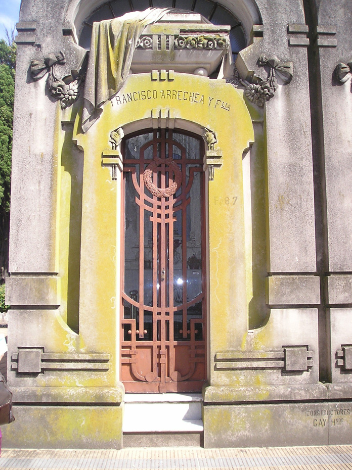
Fig. 1. Portico of the crypt of Francisco Arrechea (crypt F87), which is of architectural
(Sigma) containing 5 L of DNA and 25 pmol of each primer and
interest because of its Art Nouveau style. The yellowish coloration is indicative of
were brought to a final volume of 25 L with sterile water. PCR
the presence of Caloplaca austrocitrina.
products were analyzed by electrophoresis in 1% (wt/vol) agarose
gels in 1 × TBE buffer containing ethidium bromide (0.4 g·mL−1)
Visual inspection of crypts of historical and architectural interest
and then purified by filtration through Microcon-100 (Millipore)
showed clear evidence of significant biofouling (lichens, fungi, and
columns. Bacteria were identified by sequencing of the respective
dark biofilm patinas). Five sites from four crypts were selected for
500-bp rDNA fragment using the BigDyeTM Terminator v1.1 Cycle
Sequencing kit (L-7012, PE Applied Biosystems). Sequences were
resolved in an ABI PRISMTM 310 Genetic Analyzer following
the manufacturer's instructions and then compared directly to
crypt C95 (marble column);
all known sequences deposited in the NCBI (National Center
crypt C77 (inner side of the marble);
of Biotechnology Information) databases using the basic local
crypt F87 (front, cement mortar –
alignment search tool Megablast.
crypt A9 (front, cement mortar);
In addition, acid-forming bacteria were quantified using the
crypt A9 (side wall and front).
extinction technique, in which serially diluted cultures were main-
tained in glucose broth containing a pH indicator.
Biofilms, fungi, and crustose lichens were sampled (1 cm2) by
aseptically scraping the external surfaces of selected monuments
3. Results and discussion
using a sterile scalpel. Foliose lichens were carefully detached with
the help of a penknife. Biofilm samples to be further analyzed by
3.1. Visual inspection and biofilm analysis
scanning electron microscopy (SEM) were fixed overnight with a
2.5% glutaraldehyde solution in phosphate buffer, washed with dis-
The studied crypts were those of Vicente Isnardi (crypt C77),
tilled water, dehydrated in an acetone-water series, critical-point
Emilio B. Coutaret (crypt A9), Domingo Lastra (crypt C95), and Fran-
dried, and sputter-coated with gold. They were then examined with
cisco Arrechea (crypt F87). Vicente Isnardi was one of the founders
a Jeol scanning electron microscope (JSM-T100) at an accelerating
of La Plata University. Emilio B. Coutaret was a well-known archi-
voltage of 20 kV.
tect who assisted Pedro Benoit with his plans for La Plata Cathedral,
including the design for the Virgin's column, which is located in
the garden surrounding the Cathedral. He also built the clubhouse
of the La Plata Jockey Club. According to the La Plata Cemetery's
Lichen samples were observed by stereoscopic and optical
records, Domingo Lastra was simply an employee worker. How-
microscopy and identified using a specialized classification sys-
ever, his funerary monument is interesting not only because of its
tem to the diagnostic characteristics of the different
method of construction and the quality of the materials used, but
species; growth form, color, thallus, size, surface structures (isidia,
also because of its incomplete marble column, meant to symbolize
P.S. Guiamet et al. / Journal of Cultural Heritage 13 (2012) 339–344
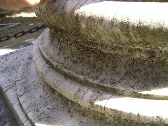
Fig. 2. Biofilm on the marble column of crypt C95.
that Lastra was unable to complete his task in this world. Francisco
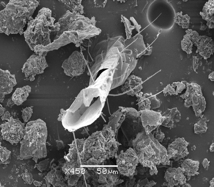
Fig. 4. Scanning electron micrograph of a sample from crypt F87, showing the pres-
Arrechea was probably a prosperous merchant or landowner. His
ence of inorganic and organic materials (fragment of an insect).
crypt is noteworthy because of its Art Nouveau style.
Lichens and dark biofilms were readily observed on crypts of
historical and architectural interest at La Plata Cemetery. Biofilms
in other monuments in La Plata, such as the Cathedral. However,
were well established on crypts with E-SE exposures, as they
other similarly constructed crypts exposed to the same conditions
receive light for only a short time during the morning and are
did not show signs of biodeterioration, most likely due to regular
exposed to the rain and humid winds coming from the La Plata
cleaning and maintenance.
River. In some cases, biofilms occurred under very specific condi-
Analysis of the biofilms by scanning electron microscopy
tions. For instance, on the column of crypt C95 (a biofilm had
revealed both fungal hyphae and bacteria embedded in a matrix
grown remarkably well on the south-facing aspect. A dark biofilm
of extracellular polymeric substances. Moreover, bdelloid rotifers
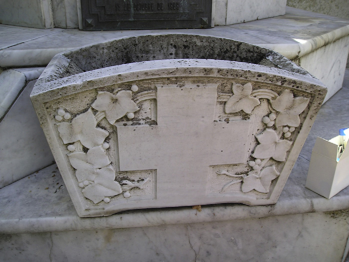
growing on crypt C77 was particularly prominent on the inner side
were also found in the crypts (Gorbushina and Petersen
of the marble flowerpot Crypt A9 was found to be in very
that the remains of arthropod exoskeletons usually
poor condition, obviously due to a lack of maintenance. Moreover,
occurred in association with growing fungi. The authors concluded
its location favored microbial colonization. Although this crypt is
that the ingestion of fungal mycelium by arthropods, as a source
situated in front of a small open square, the door faces south and
of nutrition, is accompanied by the transportation of fungal spores
the building is tucked between two other crypts, under the shade
in their intestinal tracts. Subsequent excretion of the spore mass
of a tree. The fac¸ade of crypt F87, which is covered in shade in the
results in further contamination at sites where the excreted mate-
afternoons, was colonized by lichens. The location of monuments
rial is deposited. Moreover, arthropods can directly cause substrate
in shady places facilitates the growth of lichens and other microor-
deterioration, through bioabrasion.
ganisms by reducing exposure to the sun's harmful UV rays and by
allowing the increased retention of water in the micropores of the
edifice's substrate. This was seen even in south-facing crypts and
Lichens identified as Caloplaca austrocitrina Vondrák, Riha, Arup
and Søchting Xanthoria candelaria (L.) Th. Fries, Lecanora
albescens (Hoffm) Branth and Rostr., and Xanthoparmelia farinosa
were found on cement mortar, especially on crypt F87. Approx-
imately 95% of the affected surface was covered by Caloplaca
austrocitrina and less than 5% by other lichens. Lichens belonging
to the Caloplaca genus are known to colonize natural stone. Nimis
et al. Latium and Tretiach et al. Sardinia, identified
about 300 different species of lichens on archaeological remains.
In that study, almost all Caloplaca sp. were found on calcareous
rock with a SW, S, E, or SE exposure. The exception was Caloplaca
ochracea, which occurred on northern exposures, specifically, in
shaded sites near the soil. Caloplaca was also found to have colo-
nized the carbonate rock (limestone) of the Jeronimos Monastery
(Lisbon), where lichen thalli of Thyrea, Aspicilia, and Verrucaria were
detected as well of Caloplaca sp., among others, were
also identified on the pavement mosaics of the Romanesque town
of Italica (Seville). Direct colonization of the tesserae was shown
to have been preceded by colonization of the mortar Deru-
elle et al. the nitrophiles Caloplaca citrina, Xanthoria
Fig. 3. Dark biofilm on a marble flowerpot across from crypt C77.
candelaria, and Lecanora albescens on the Basilica of Notre-Dame de
P.S. Guiamet et al. / Journal of Cultural Heritage 13 (2012) 339–344
Microorganisms found in various crypts at La Plata Cemetery (Argentina).
Total aerobic bacteria
Acid-forming bacteria
Crypt C95 (marble column)
Crypt C77 (marble flower pot, internal side)
Crypt A9 (side wall)
L'Epine (a calcareous monument in Marne, France). The presence of
also identified by Wollenzein et al. the causative agents of
these species indicated that nutrient enrichment of the stone sub-
the deterioration of marble and other calcareous materials used
strate had favored the lichens' growth. Overall, the recent increase
in the building of culturally important monuments in many local-
in lichen colonization may be related to the widespread use of fer-
ities in the Mediterranean area. Gaylarde and Gaylarde in a
tilizers and above all to the newly implemented method of spraying
comparative study of the microbial biomass of biofilms occurring
by pulverization. This was reflected by the broader extent of lichen
on the exteriors of buildings in Europe and Latin America, found
populations, augmenting the disfigurement on the west-facing sur-
that fungi, although not an important contributor to the biomass
faces, which are more exposed to the dominant winds.
invading stone, seemed to preferentially colonize painted surfaces
Bech-Andersen and Christensen that in cement-
rather than other substrates (cement, mortar, concrete). Perfettini
based materials the cement paste was preferentially attacked by
et al. a strain of Aspergillus that produced gluconic and
lichens, leaving the aggregates unaffected. More recent studies
oxalic acids during the degradation of cement. After 8 months of
showed that Caloplaca austrocitrina can penetrate both cement and
contact with the substrate, these acids had induced the dissolution
concrete. This species also produces oxalic acid, whose chemical
of portlandite (without the leaching of calcium) and had increased
action causes a loss of calcium from mortar substrates a
the porosity of the cement while reducing its bending strength.
study of stone monuments in Rome, Seward the dif-
In more recent laboratory studies, ordinary Portland cement
ferential actions of lichen species in biodeterioration. Furthermore,
pastes were severely attacked during bioleaching using Aspergillus
not all species are harmful. While the sporadic growth of Lecanora
niger cultures, especially by fungal biogenic organic (acetic, butyric,
muralis has been related to the mechanical decay of the substrate,
lactic, and oxalic) acids and to a lesser extent by respiration-
species such as Lecanora dispersa, which also account for significant
induced carbonic acid However, this study was conducted
coverage, do not cause evident modifications. Monte
under atypical conditions, in which large amounts of water were
that Lecanora campestris and Lecanora rupicola preferentially grow
present, and cannot be related to the conditions to which most
on W to SW exposures whereas Xanthoria calcicola grows mainly
buildings are exposed.
on south-facing surfaces. These preferences are consistent with
The turgor pressure exerted by the hyphae of plant pathogens
the fact that some species are stenoic, thriving only within a very
such as the rice blast fungus Magnaporthe grisea has been calculated
specific range of pH, humidity, luminosity, and nitrogen supply,
on synthetic surfaces, including the plastic poly(vinyl chloride)
while others are euroic and thus tolerant of a wider range of condi-
measurement of turgor pressure indicated that the
tions their role as primary colonizer seems doubtful,
hyphae of this fungus can generate pressures in excess of 8.0 MPa,
lichens nonetheless cause reduced substrate cohesion and there-
which is sufficient to allow fungal penetration of marble (tension
fore biocorrosion. In the crypts sampled in the present study, the
resistance of 3.9 MPa), thereby causing mechanical damage at the
surfaces were rough, eroded, and uneven due to the chemically
targeted destruction of the cement matrix but not the aggregates,
which were unaffected. Mechanical damage, by contrast, results
from penetration of the substrate by lichen hyphae. The penetration
depth of the thallus depends on the lichen species and the nature of
Total aerobic bacteria and acid-forming bacteria counts are
the substrate may also cause substrate damage by the
shown in Based on these counts, cement mortar was
production of acidic substances, such as carbon dioxide, lichenic
more highly colonized than marble. From the isolated bac-
acids, and oxalic acid. A full review of lichens as agents of damage
teria, three isolates were selected for sequencing based on
can be found in Lisci et al.
the Gram staining and API results. These bacteria were sub-
While chemolithotrophic microorganisms have often been
sequently identified as Bacillus sp. and Pseudomonas sp. Their
described in association with damaged inorganic materials, more
sequences were deposited in GenBank under the accession num-
recent studies have emphasized the significance of chemoorgan-
bers JN837477 (97% similarity with JN208185), JN837478 (98%
otrophic bacteria and fungi, together with photoautotrophs, as
similarity with HM352366), and JN837479 (96% similarity with
the primary colonizers of building stones. The activities of
AY269246). Bacillus sp. (spore-forming bacteria) and Pseudomonas
these primary colonizers precondition the building for attack by
sp. (desiccation-tolerant bacteria) are able to survive in non-
chemolithoautotrophs and thus initiate the process of biological
favorable conditions and can be easily found in environmental
Heterotrophic bacteria have frequently been isolated on stone
monuments and are known to cause biodeterioration
et al. several species of the genera Bacillus on weathered
Aspergillus sp. and Penicillium sp. were the predominant groups
sandstones of a church. Vuorinen et al. that Finnish
probably because they are ubiquitous air-borne fungi and easily
granite is slowly degraded by cultures of Pseudomonas aeruginosa,
cultivable. Fusarium sp., Candida sp., and Rhodotorula sp. were also
resulting in morphological alteration of the stone surface and the
detected among the fungi that colonized La Plata Cemetery's mar-
elution of minerals from the stone.
ble and cement mortar crypts. The fungal-mediated deterioration
In the crypts examined in this study, acid-forming bacteria
of stone was previously reviewed by May et al. were
were isolated from marble and from cement mortar. These bacteria
P.S. Guiamet et al. / Journal of Cultural Heritage 13 (2012) 339–344
are likely to play a role in the biodeterioration of these historical
the material (discoloration, salt efflorescences, etc.) and should be
monuments through the chemical and physical processes that are
specific for the biodeteriorating organisms but not harmful to peo-
integral to biofilm formation
ple, other animals, or plants.
3.5. Bioreceptivity of the building materials
According to Guillitte describes a material
Mature biofilms cover crypts of historical and architectural
that can be colonized by living organisms but without neces-
interest at La Plata Cemetery (Argentina). These biofilms have
sarily undergoing biodeterioration. The bioreceptivity of stone is
caused the biodeterioration of monuments of different periods,
determined by its structure and chemical composition, and the
styles, and materials.
intensity of microbial contamination by climatic conditions and
Lichens (Caloplaca austrocitrina, Lecanora albescens, Xan-
anthropogenic eutrophication of the atmosphere
thoparmelia farinosa, Xanthoria fallax), fungi (Aspergillus sp.,
given the same climate, the influence of the substrate on micro-
Fusarium sp., Penicillium sp., Candida sp., Rhodotorula sp.), and
bial colonization can be examined. Interestingly, our data (
bacteria (Bacillus sp., Pseudomonas sp.) were identified based on
show that in spite of similarities in microclimate, microbial growth
morphological and biochemical characteristics and in the case of
on marble and cement mortar differed. While the cement mor-
bacteria by rDNA sequencing as well. These organisms are not a
tar was almost completely covered by lichens, the marble was
complete list of those present on the crypts. Phototrophs (algae
less colonized. In addition, bacterial and fungal growth on cement
and cyanobacteria) were not sought, but there is no doubt that
mortar was greater than on marble. A possible explanation for
they are present as primary colonizers of building stones. The
this is that cement mortar has a higher porosity than marble. In
use of PCR-based molecular tools on fresh biofilms, rather than
laboratory tests performed at the LEMIT ("Laboratorio de Entre-
culture-dependent techniques, would have allowed many more
namiento Multidisciplinario para la Investigación Tecnológica", La
microorganisms to be detected.
Plata, Argentina), the water absorption rate of samples of Car-
rara marble was 0.924% (indicating a low porosity), whereas that
of cement mortar was 6–9% (indicating a high porosity). Thus,
regardless of the heterogeneity of cement mortars in terms of their
The authors thank the "Universidad Politécnica de Madrid"
composition, cement/water relationship, and age, they are much
(Spain) for its financial support (grants AL07-PID-020 and AL08-
more porous than marble. This ability to absorb and retain rela-
P (I + D)-08). P.S. Guiamet, V. Rosato, and S. Gómez de Saravia
tively large amounts of water provides favorable growth conditions
gratefully acknowledge the "Consejo Nacional de Investigaciones
for microorganisms.
Científicas y Técnicas" (CONICET) and the "Universidad Nacional
de La Plata" (UNLP) "Proyecto de Incentivos" 11 N 578.
3.6. Conservation and restoration of crypts
Some authors are of the opinion that if biofilms do not cause
serious damage or hinder conservation efforts then their presence
[1] J.A. Morosi, La Plata an advanced nineteenth century new town with ancient
may actually increase the cultural value of a building or monu-
roots, Plan Perspect. 18 (2003) 23–46.
ment However, this is not the case at La Plata Cemetery,
[2] D. Allsopp, K. Seal, C.C. Gaylarde, Introduction to Biodeterioration, 2nd edition,
where biodeterioration of the crypts is serious enough to warrant
Cambridge University Press, Cambridge, 2004.
[3] P. Ozenda, G. Clauzade, Les Lichens. Étude biologique et flore illustreé, Ed.
Masson et Cie, Paris, France, 1970 (in French).
The removal of lichens and other organisms from a surface is a
[4] J. Poelt, Bestimmungschlüssel europäischer Flechten, Ed. J. Cramer, 1969 (in
delicate task that must be considered and planned with the great-
est care. It differs from a simple cleaning, which allows regrowth.
[5] K.H. Domsch, W. Gams, T.-H. Anderson, Compendium of Soil Fungi, Academic
Press, London, 1993.
In Buenos Aires Province, for example, regrowth of the lichen Calo-
[6] A.A. Gorbushina, K. Petersen, Distribution of microorganisms on ancient wall
placa austrocitrina on the region's stone monuments and cement
paintings as related to associated faunal elements, Int. Biodeterior. Biodegrad.
mortar-covered buildings was observed 6 months after these struc-
46 (2000) 277–284.
[7] V.G. Rosato, U. Arup, Caloplaca austrocitrina (Teloschistaceae) new for South
tures had been cleaned using a hydrojet washing method (our
America, based on molecular and morphological studies, The Bryologist 113
unpublished results). Lichen reproduction occurs by the germina-
(2010) 124–128.
tion of spores or by multiplication of the cells of the soredia and
[8] P.L. Nimis, M. Monte, M. Tretiach, Flora e vegetazione lichenica di aree arche-
ologiche del Lazio, Stud. Geobot. 7 (1987) 3–161 (in Italian).
isidia. These reproductive structures are dispersed in the atmo-
[9] M. Tretiach, M. Monte, P.L. Nimis, A new hygrophytism index for epilithic
sphere by wind or transported by birds and insects. If they settle
lichens developed on basaltic Nuraghes in NW Sardinia, Bot. Chron. 10 (1991)
into cracks, pores, or cavities that retain water, new growth may
[10] C. Ascaso, J. Wierzchos, V. Souza-Egipsy, A. de los Ríos, J. Delgado Rodrigues,
arise. On stone surfaces, the microorganisms comprising the lichen
In situ evaluation of the biodeteriorating action of microorganisms and the
thallus are frequently accompanied by bacteria, cyanobacteria,
effects of biocides on carbonate rock of the Jeronimos Monastery (Lisbon), Int.
free-living algae, or fungi, and their additional effects cannot be
Biodeterior. Biodegrad. 49 (2002) 1–12.
[11] J. Garcia Rowe, C. Saiz-Jimenez, Colonization of mosaics by lichens: the case
ignored in any removal strategy
study of Italica (Spain), Stud. Geobot. 8 (1988) 65–71.
Before a decision on the removal method is reached and a bio-
[12] S. Deruelle, R. Lallemant, C. Roux, La végétation lichenique de la Basilique Notre-
cide is chosen, the environmental conditions (for example, light
Dame de L'Epine (Marne), Documents Phytosociologiques 4 (1979) 217–234 (in
and humidity) must be considered and, to the degree possible,
[13] J. Bech-Andersen, P. Christensen, Studies of lichen growth and deterioration
changed; otherwise, the effects of the treatment will be short last-
of rocks and building materials using optical methods, in: T.A. Oxley, S. Barry
ing. Unfortunately, this is not possible in the case of the La Plata
(Eds.), Biodeterioration 5, John Wiley & Sons Ltd, 1983, pp. 568–572.
funerary monuments, since they are regularly exposed to inclement
[14] L.P. Traversa, S. Zicarelli, R. Iasi, V.G. Rosato, Biodeterioro de morteros y
hormigones por acción de los líquenes, Hormigón 35 (2000) 39–48 (in Spanish).
weather. The nature of the material, the degree of its alteration,
[15] M.R.D. Seaward, Lichen damage to ancient monuments: a case study, Lichenol-
and the presence and density of the organisms causing the biode-
ogist 20 (1988) 291–294.
terioration are critical factors in deciding whether cleaning and
[16] M. Monte, The influence of environmental conditions on the reproduction and
distribution of epilithic lichens, Aerobiologia 9 (1993) 169–179.
removal are required and whether a biocide will be needed
[17] M. Lisci, M. Monte, E. Pacini, Lichens and higher plants on stone: a review, Int.
authors of the latter study emphasized that biocides must not alter
Biodeterior. Biodegrad. 51 (2003) 1–17.
P.S. Guiamet et al. / Journal of Cultural Heritage 13 (2012) 339–344
[18] C. Gehrmann, W.E. Krumbein, K. Petersen, Lichen weathering activities on min-
[25] R.J. Howard, M.A. Ferrari, D.H. Roach, N.P. Money, Penetration of hard substrates
eral and rock surfaces, Stud. Geobot. 8 (1988) 33–45.
by a fungus employing enormous turgor pressures, Proc. Natl. Acad. Sci. USA
[19] S. Scheerer, O. Ortega-Morales, C. Gaylarde, Microbial deterioration of stone
88 (1991) 11281–11284.
monuments: an updated overview, Adv. Appl. Microbiol. 66 (2009) 97–139.
[26] E. Book, W. Sand, The microbiology of masonry biodeterioration: a review,
[20] E. May, F.J. Lewis, S. Pereira, S. Tayler, M.R.D. Seaward, D. Allsopp, Micro-
J. Appl. Bacteriol. 74 (1993) 503–514.
bial deterioration of building stone: a review, Biodetererior. Abstr. 7 (1993)
[27] M. Flores, C. Fernandez, M. Barbáchano, Microbial biodeterioration of stone
in historic-artistic monuments, in: S.A.C. Sequeira, A.K. Tiller (Eds.), Microbial
[21] U. Wollenzien, G.S. de Hoog, W.E. Krumbein, C. Urzi, On the isolation of micro-
Corrosion, The Institute of Materials, London, 1992, pp. 262–265.
colonial fungi occurring on and in marble and other calcareous rocks, Sci. Total
[28] A. Vuorinen, S. Mantere-Alhonen, R. Uusinoka, P. Alhonen, Bacterial weathering
Environ. 167 (1995) 287–294.
of Rapakivi granite, Geomicrob. J. 2 (1981) 317–325.
[22] C.C. Gaylarde, P.M. Gaylarde, A comparative study of the major microbial
[29] O. Guillitte, Bioreceptivity: a new concept for building ecology studies, Sci. Total
biomass of biofilms on exteriors of buildings in Europe and Latin America, Int.
Environ. 167 (1995) 215–220.
Biodeterior. Biodegrad. 55 (2005) 131–139.
[30] Th. Warscheid, J. Braams, Biodeterioration of stone: a review, Int. Biodeterior.
[23] J.V. Perfettini, N. Langomazino, E. Revertegat, J. Petit, Evaluation of the cement
Biodegrad. 46 (2000) 343–368.
degradation induced by the metabolic products of two fungal strains, in: C.C.
[31] G. Caneva, M.P. Nugari, O. Salvadori, La biologia vegetale per i beni cul-
Gaylarde, L.H.G. Morton (Eds.), Biocorrosion, Proceedings of a Joint Meet-
turali, Vol. 1: Biodeterioramento e conservazione, Nardini, Florencia, 2005
ing between the Biodeterioration Society and the French Microbial Corrosion
(in Italian).
Group, Paris, France, 1989, pp. 23–36.
[24] L. de Windt, P. Devillers, Modeling the degradation of Portland cement pastes
by biogenic organic acids, Cem. Concr. Res. 40 (2010) 1165–1174.
Source: http://lemac.frlp.utn.edu.ar/wp-content/uploads/2012/09/2012_Cementerio-La-Plata_Journal-Of-Cultural-Heritage.pdf
N e w s l e t t e r vol. 10, no. 4 - 2007 "the recent initiative of EU Commission to identify "lead markets for biobased products" has shown that there is a need for realistic surveys in the EU-markets for RRMs and RRM based products.In the last edition of Green Tech letters 3/2007 the French Agency ADEME published the results of the ALCIMED survey on existing markets and future perspectives in France.
Flexible Spending Accounts Eligible Expense Guide Healthcare & Dependent Care Eligible Expense Guide This guide will provide a detailed listing of a healthcare and dependent care FSA spending account. It lists general expenses allowed by the Internal Revenue Service (IRS), however, it is not an exhaustive list and if you have a question regarding an expense not listed, please call the RPG Consultants FSA Department at (212) 947-4800 ext. 215. How to use this guide 1. Healthcare & Dependent Care: Sections contain an easy-to-read chart listing common expenses. 2. Expense Type: Expenses are displayed alphabetically within their category. Examples of the expense are listed beneath the expense. 3. Eligible for Reimbursement: This guide will state if the expense is generally reimbursable from your FSA. However, there are certain exceptions or requirements for many expenses. It is important that you read the Special Exceptions or Requirement related to the expense. 4. Special Exceptions or Requirements: Follow the instructions provided to ensure your particular expense is eligible. Tip: To quickly search for an expense, click the search icon and type in the item You are looking for. Example: You're searching for glasses, just type in ‘eye glasses.' General rules for eligible expenses In order for RPG Consultants to consider an expense eligible, the expense must: • Be allowed by the IRS; • Be allowed by your employer's FSA plan; • Be incurred during your employer's plan year or during the 2 ½ month extension period, if allowed by your employer's plan; • Be for you or a qualified dependent • Not be reimbursed through another plan • Not be used as a dependent day care credit or healthcare deduction on your personal tax return. To obtain reimbursement for an eligible healthcare or dependent care expense, you must complete a reimbursement request, which can be found on our web site www.rpgny.com and must be accompanied by a detailed receipt or other supporting documentation. Definition of a qualifying dependent Healthcare and dependent care expenses must be for you or a qualified dependent in order to be eligible for your FSA. The definition of "dependent." Please note: Generally speaking, a qualifying child must: • Reside with you for more than half the year1; • Be younger than 27 years of age • Not provide over half of his/her own support.


