Medical Care |

Medical Care

##SEVER##

//p/pt.armstrong.edu1.html


Untitled

[ RESIDENT'S CASE PROBLEM ]
MICHAEL S. CROWELL, PT, DPT¹šDEHC7DM$=?BB" PT, DSC, OCS, FAAOMPT²
Medical Screening and Evacuation: Cauda Equina Syndrome in a Combat Zone Low back pain (LBP) is a prevalent condition, particularly in cific LBP, nerve root syndrome (radicu-
primary care clinics, with billions of dollars spent each year lopathy or stenosis), and serious spinal on treatment.33 It is the fifth most common reason for all LBP secondary to nerve root syn- physician visits in the United States.11 Approximately 25% of drome, although less common, is a po- adults report LBP lasting at least 1 day within the past 3 months,11 tentially disabling condition.23 Nerve with approximately 14% having an episode that lasts longer than 2 root syndrome may be related to radicu- weeks.15 Prevalence ranges from 15% to 20% over a single year40 lopathy, spinal stenosis, or cauda equina and approximately 70% over the course of a person's lifetime.15,27,30,40 syndrome (CES).23 Due to the potentialfor poor prognosis, timely recognition of The majority of patients (85%) ease or spinal abnormality.11,15 Current neurologic involvement is essential for with LBP have conditions that cannot recommendations suggest classifying optimal patient outcomes.23 be reliably attributed to a specific dis- patients into 3 broad categories: nonspe- Acute lumbar disc herniation is one potential source of both radiculopathy TIJK:O:;I?=D0 Resident's case problem.
weakness, absent right ankle reflex, and decreased and CES. Approximately 90% of cases ofsciatica are caused by a herniated disc.29 anal sphincter tone. No advanced medical imaging Cauda equina syndrome (CES) Overall incidence of symptomatic disc is a rare, potentially devastating, disorder and is capabilities were available locally. Due to suspected considered a true neurologic emergency. CES often CES, the patient was medically evacuated to a neuro- herniation is 1% to 2%,29,39 for which has a rapid clinical progression, making timely rec- surgeon and within 48 hours underwent an emergent 200 000 discectomies are performed an- ognition and immediate surgical referral essential.
L4-5 laminectomy/decompression. He returned to full nually.39 Peak incidence of this disorder is military duty 18 weeks after surgery without back or T:?7=DEI?I0 A 32-year-old male presented to a
between the ages of 30 to 55 years.15 lower extremity symptoms or neurological deficit.
medical aid station in Iraq with a history of 4 weeks Characteristics of acute disc hernia- of insidious onset and recent worsening of low back, T:?I9KII?ED0 This case demonstrates the im-
tion include abrupt, intense onset of left buttock, and posterior left thigh pain. He denied portance of continual medical screening for physi- pain that is increased by bending or lift- symptoms distal to the knee, paresthesias, saddle cal therapists throughout the patient management ing.48 The most common levels of symp- anesthesia, or bowel and bladder function changes.
cycle. It further demonstrates the importance of tomatic herniation are L5-S1 and L4-5, At the initial examination, the patient was neuro- immediate referral to surgical specialists when logically intact throughout all lumbosacral levels CES is suspected, as rapid intervention offers the which comprise approximately 90% to with negative straight-leg raises. He also presented best prognosis for recovery.
98% of cases11,15 and correspond to the with severely limited lumbar flexion active range of spinal levels that receive the majority of motion, and reduction of symptoms occurred with level 4. J Orthop Sports Phys Ther 2009;39(7):541- compressive forces in the lumbar spine.
repeated lumbar extension. At the follow-up visit, 10 549. doi: 10.2519/jospt.2009.2999 Clinically, disc herniation at these levels days later, he reported a new, sudden onset of saddle frequently manifests as L5 and S1 nerve anesthesia, constipation, and urinary hesitancy, TA;OMEH:I0 direct access, lumbar spine, low
root compression disorders character- with physical exam findings of right plantar flexion back pain, red flags, spinal cord ized by radiating pain below the knee, 1Brigade Combat Team Physical Therapist, Iraq. 2Program Director and Associate Professor, US Army-Baylor University Postprofessional Doctoral Program in Orthopedic ManualPhysical Therapy, Brooke Army Medical Center, Fort Sam Houston, TX. This case was seen at a Troop Medical Clinic in Iraq. The opinions or assertions contained herein are theprivate views of the authors and are not to be construed as official or reflecting the views of the United States Army or Department of Defense. Address correspondence to CPTMichael Crowell, 235 Lancaster Way, Richmond Hill, GA 31324. E-mail: [email protected] journal of orthopaedic & sports physical therapy volume 39 number 7 july 2009 541 [ RESIDENT'S CASE PROBLEM ]
History of Present Illness
Unrelenting night pain History of cancer or recent infection Unexplained weight loss or gain The patient was a 32-year-old
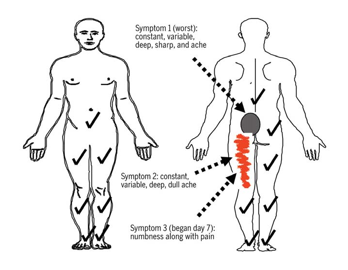
Caucasian male (height, 1.83 m;body mass, 77.1 kg; body mass in- dex, 23.1 kg/m2) who initially presented to a physical therapist in a combat zone Difficulty with micturation* with a chief complaint of insidious onset Loss of anal sphincter tone or fecal incontinence* low back and left posterior thigh pain. He Saddle anesthesia* was deployed for combat with a primary Gait disturbance* responsibility of training foreign military * Indicates elements specific to cauda equina syndrome. officers. During convoy operations, he wasa machine gunner, standing in the turret decreased sensation in a dermatomal embryo development, the spinal cord and at the top of an armored vehicle. This job pattern, myotomal weakness, reflex vertebral column have relatively unequal required prolonged periods of wearing changes, and positive straight-leg raise rates of growth.20 As a result, the lumbar protective equipment weighing in ex- tests.11,15 However, examination of pa- and sacral spinal nerves descend almost cess of 36 kg, often for periods exceed- tients with acute disc herniation should vertically to reach their points of exit.20 ing 8 hours. His symptoms were located always include careful screening for se- This configuration resembles a "horse's in the lower lumbar spine (left greater rious pathology, both before initiation tail," from which the term cauda equina than right), left buttock, and left poste- of and during ongoing conservative is derived in Latin.
rior thigh, as shown in <?=KH;'. The total
interventions.3,11 Red flag differential CES is a rare, potentially devastating duration of symptoms was approximately diagnoses may include CES, metastatic disorder that may arise from an acute 4 weeks, but the patient reported a sig- spinal disease, spinal infection, epi- disc herniation and is considered a true nificant increase in intensity of pain the dural hematoma, and spinal fracture neurologic emergency.8,45,48 The estimated day prior to evaluation without any spe- or dislocation.3 Screening for red flag prevalence of CES is 0.04% of all patients cific trauma. He described a dull, aching conditions should include questions presenting with a primary complaint of pain, and a pain that was intermittently regarding bowel and bladder function LBP,11,15,45 and it is most prevalent in the sharp. The patient denied numbness or changes, sensory function changes in fourth or fifth decade of age.43,45 CES oc- tingling in any location or pain below the the perianal region and genitals, unex- curs in 1% to 2% of all lumbar disc her- level of the knee. Baseline numeric pain plained weight loss or gain, fever, night niations that progress to surgery.1,8,15,43,44 rating scale (NPRS),25 where 0 is no pain pain, history of cancer or infection, CES is most frequently associated with and 10 is the worst pain that the patient history of trauma, and any gait distur- a nontraumatic massive midline posteri- could imagine, was 4/10 at rest and 7/10 bances (TABLE 1).3,22
or disc herniation, commonly located at at worst. The patient noted increased In adults, the spinal cord is approxi- L4-5, followed by L5-S1 and L3-4.43,44,45,48 pain with running and forward flexion of mately 42 to 45 cm in length and ter- The sacral nerves, which lie medially in the lumbar spine. Rest and lying supine minates at the lower border of the first the cauda equina, are affected dispropor- relieved his symptoms. His past medical lumbar vertebra or upper border of the tionately in this disorder. A clear diagno- history was significant for 3 to 4 prior oc- second lumbar vertebra at the conus med- sis or a high index of suspicion for CES currences of LBP over the past 8 years, ullaris.20 The spinal cord is ensheathed by should prompt immediate referral to a with similar presentation that, he stated, 3 protective membranes from outward to surgical specialist. Referral to an ortho- resolved without treatment. The patient within: the dura mater, arachnoid, and paedic spine surgeon or neurosurgeon, had no history of spine or extremity sur- pia mater.20 These membranes extend where available, is the most direct route gery. No previous imaging studies had to the first segment of the coccyx as the of referral; otherwise, the patient should been performed. His stated goal was to filum terminale.20 The outer layer of the be sent to an emergency department.
decrease his overall pain level during per- dura, arachnoid, and the subarachnoid The purpose of this resident's case formance of his military duties.
cavity is termed the thecal sac, which is problem is to describe the evaluation, filled with cerebrospinal fluid.20 treatment, referral, and outcomes of a The term cauda equina describes the patient exhibiting signs and symptoms The patient denied saddle anesthesia, lumbar and sacral spinal nerves descend- of CES evaluated by a physical therapist bowel or bladder function changes, unex- ing from the conus medullaris. During in a direct-access environment.
plained weight loss or gain, night pain, or 542 july 2009 volume 39 number 7 journal of orthopaedic & sports physical therapy


Assessment, Intervention,
and Re-evaluation
Using a treatment-based classification
approach,6,7,18 the patient was classified
into the specific exercise classification
and prescribed extension-oriented exer-
cises. Either standing or prone repeated
extension exercises were to be performed
every 2 waking hours, with 10 repetitions,
holding each repetition 2 to 3 seconds at
end range. Education consisted of avoid-
ance of sitting for greater than 20 to 30
minutes, avoidance of full end-range
flexion positions, and the use of a lumbar
roll while sitting and wearing protective
equipment. The treating physical thera-
pist, who had privileges for prescribing
nonnarcotic medication,5 prescribed 7.5
mg Meloxicam (Mobic, 2 tablets once
daily) and 500 mg acetaminophen (Tyle-
nol, 2 tablets every 4-6 hours, as needed)
for pain relief during performance of his
<?=KH;'$Body chart of symptoms at initial presentation through day 9.
military duties. Because this patient was recent trauma, and had no history of cancer back, left buttock, and left posterior thigh essential to the success of his unit's mis- or infection. In screening for nonmusculo- at the end range of motion. Lumbosacral sion, only a home exercise program was skeletal pathology, the patient reported no extension and side bending were within prescribed, and a follow-up was sched- history of cancer, cardiovascular, or pul- normal limits, without an increase in uled for 2 weeks later. He was instructed monary disease, and no recent occurrence pain from his baseline NPRS. Repeated- to return to the clinic at an earlier time of nausea, vomiting, fever, changes in ap- motion testing was performed as de- for re-evaluation if symptoms worsened.
petite, difficulty swallowing, shortness of scribed by McKenzie.35 Ten repetitions The patient presented for follow-up on breath, dizziness, or changes in balance.
of flexion in standing increased his back, day 4 (3 days after the initial evaluation) buttock, and posterior thigh pain, while with a complaint of increased pain unre- Test and Measures
10 repetitions of extension in standing lieved by positioning and only short-term The patient was neurologically intact reduced those symptoms. Straight-leg relief with the home exercise program.
bilaterally with 5/5 strength as assessed raise tests did not produce radicular pain His baseline NPRS at rest had increased with manual muscle testing26 through- but caused a severe increase in LBP at 15° to 6/10. He reported a decreased NPRS out the L2 to S1 myotomes, sensation of hip flexion on the right and 45° of hip to 4/10 after performing home exercises, was intact to light touch throughout the flexion on the left. Hip flexion range of but he would return to baseline after ap- L2 to S1 dermatomes, and knee jerk and motion during single knee to chest was proximately 30 to 60 minutes. Since the ankle jerk muscle stretch reflexes were within normal limits bilaterally, with in- initial evaluation, he had continued to 2+ (normal). Babinski reflex testing was creased LBP that was approximately 50% perform all of his duties, including ex- negative. He presented with decreased less than with straight-leg raise testing.
tended wear of protective equipment.
lumbar lordosis and a guarded, obviously A lumbar quadrant test was negative bi- The physical examination, including the painful, movement of the spine. The pa- laterally. Passive vertebral motion testing, neurological examination, did not differ tient displayed a left-sided antalgic gait, as described by Maitland,34 produced lo- from the initial evaluation, with the ex- with decreased left hip extension and cal pain at L3, L4, and L5 with central ception of increased pain with all testing.
early termination of the stance phase of passive posterior-anterior accessory in- His case was discussed with a physician the gait cycle. Active range of motion was tervertebral motion (PAIVM). Unilateral and he was prescribed a narcotic pain severely limited in lumbosacral flexion, PAIVM testing produced local pain at medication for use as needed, re-educat- with the ability to reach only the mid- L3-4, L4-5, and L5-S1, equal bilaterally.
ed in the home exercises to ensure proper anterior thigh region with the fingertips, No referral of pain was noted with pas- performance, and instructed to continue and moderate to severe pain in the low sive accessory movement assessment.
the home exercises as tolerated. The pa- journal of orthopaedic & sports physical therapy volume 39 number 7 july 2009 543


[ RESIDENT'S CASE PROBLEM ]
tient was to follow-up within 1 week tomonitor the stability of his symptoms andassess his response to the modified treat-ment plan.
At a second follow-up 3 days later (day 7 after initial exam), the patient contin-ued to have significant pain. He was ableto perform his duties, but the sharp painwas becoming more frequent and in-tense. He also reported a recent onsetof numbness in his left posterior thigh.
He continued to deny any radiating painbelow the knee or right-sided symptoms.
The physical exam was unchanged frominitial evaluation and a neurological as-sessment continued to reveal no mo-tor, sensory, or reflex deficits bilaterally.
Although strength of left ankle plantarflexion was 3+/5, he was limited by painsecondary to a recent ankle inversionsprain on rocky, uneven terrain, which <?=KH;($Body chart of symptoms at 10-day follow-up.
he described as unrelated to his low backsymptoms and had occurred between the holds, with 3 repetitions. The patient's evaluation. A straight-leg raise test bilat- first and second re-evaluation.
home exercise program remained un- erally continued to provoke symptoms Due to increasing pain despite conser- changed from the initial evaluation.
only in the low back region. No sensory vative therapy and medications, he was Upon presenting for his third day of deficiencies to light touch or sharp-dull restricted from missions that required the in-clinic treatment (10 days after initial stimuli were noted throughout the lower wear of his protective gear and from any evaluation), the patient had a new com- extremities bilaterally, including the L4- lifting, bending, or twisting. Daily physi- plaint of numbness and tingling in the S1 dermatomes. Strength was reduced in cal therapy intervention in the clinic was saddle region and a change in bowel and right ankle plantar flexion to 3–/5. The initiated at that time. The therapist chose bladder function. Although he denied any right ankle jerk (S1) reflex was absent. A to continue with a supervised exercise incontinence, he stated that it was diffi- rectal examination revealed decreased program and adjunct pain-relieving mo- cult to control initiation and cessation anal sphincter tone and an absent anal dalities, because the high-load demands of urination and bowel movements. The wink reflex. The cremasteric reflex was of this patient's work duties up to that patient also described new symptoms intact. The Babinski reflex was normal.
point made accurate assessment of the in the right lower extremity (previously Gait was severely impaired with a de- patient's response to treatment difficult.
asymptomatic), with an inability to rise creased step length bilaterally and im- Intervention consisted of interferential up onto his toes and constant tingling in paired toe-off present on the right.
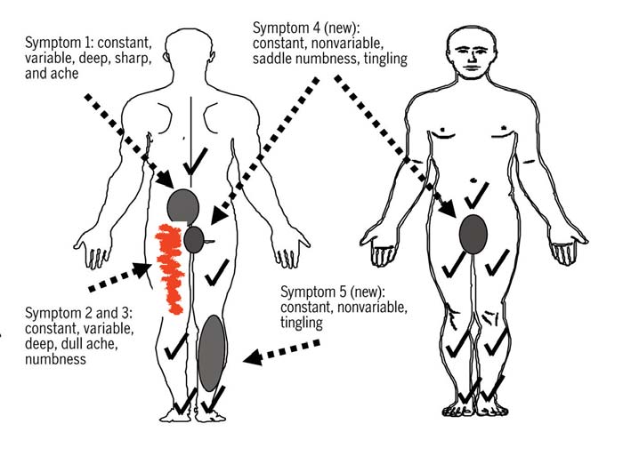
electrical stimulation with 4 pads brack- the right calf, while his left lower extrem- eting the symptomatic area of the lumbar ity symptoms were unchanged from the spine, the patient positioned in prone, last evaluation. <?=KH;( shows the body
Because of his rapidly progressive neu- and the intensity at the patient's level of chart associated with the new symptom rological symptoms and a suspicion of tolerance. Treatment was combined with presentation. He stated, however, that his cauda equina compression, the physi- moist heat for 20 minutes, followed by pain level had decreased to 4/10 at rest cal therapist scheduled the patient for supervised extension exercises and left and 5/10 at worst since he stopped wear- medical evacuation and referral to a lumbar rotation stretches, both of which ing his protective gear 2 days prior.
neurosurgeon. No advanced imaging was provided mild relief of the lower extrem- A detailed physical examination was performed, as magnetic resonance imag- ity pain. Lumbar extension exercises con- performed, with an orthopaedic physi- ing (MRI) and computed tomography sisted of 3 sets of 10 repetitions, with a cian assistant on staff providing further (CT) scan capabilities were not available 2- to 3-second hold at end range, without guidance on neurological assessment of at the local facility. Evacuation to neuro- manual overpressure. Lumbar left rota- the S3-4 levels. Lumbar spine range of surgery care and advanced medical imag- tion stretching consisted of 30-second motion was unchanged from the initial ing occurred within 48 hours.
544 july 2009 volume 39 number 7 journal of orthopaedic & sports physical therapy ing 1.6 km daily, doing pool exercises at Radiology Impression of Computed Tomography home, and using 1- to 2-kg weights for (CT) Scan Prior to Surgical Intervention upper extremity exercises. At 4-monthfollow-up he had no residual neurological L1-2: No disk bulge, central canal, or neuroforaminal stenosis or functional deficits and reported a cur- L2-3: No disk bulge, central canal, or neuroforaminal stenosis rent walking program with a 5-kg back- L3-4: Broad-based disk bulge with no sadistic and central canal or neural foraminal stenosis pack. He was cleared by neurosurgery for L4-5: Moderate disk space narrowing; degenerative changes of the inferior endplate of L4; large disk protrusion with full return to military duties, including moderate central canal stenosis, difficult to tell, but likely some extruded fragments posterior to L5; moderate to severe lateral recess stenosis bilaterally; exiting nerve roots in the neural foramina appear relatively normal L5-S1: No significant disk bulge, central canal, or neural frontal stenosis and, to enhance functional recovery, with lower extremity exercises aimed to main- Upon arrival at a Combat Support Hos- tain nervous tissue mobility to prevent This resident's case problemde-
scribes what could be considered a pital, the patient was evaluated by a mili- postoperative nerve root scarring. After classic presentation of CES, recog- tary neurosurgeon. Physical examination 3 days in Germany, the patient was then nized by a physical therapist practicing findings were consistent with those at the evacuated to his final destination, Brooke in a direct-access setting. By a continual medical aid station. Additionally, bladder Army Medical Center, Fort Sam Houston, medical-screening process over multiple function was evaluated and the patient TX, for follow-up neurosurgical care and visits, the therapist recognized an atypi- was found to have a postvoid residual cal progression of mechanical LBP, which of 300 cc. Although MRI is the recom- then acutely manifested itself as CES.
mended imaging modality for CES, be- Early recognition, confirmation, referral, cause of the detail provided to the soft The patient arrived at Brooke Army Med- and surgical intervention were associated tissues and spinal canal,11 it was not avail- ical Center 6 days after surgery. An MRI with a good outcome, consistent with lit- able in that location either. Instead, a CT performed on admission demonstrated erature that suggests a good prognosis of the lumbar spine with contrast, an al- normal postoperative changes and no with early detection and treatment.1,43,44 ternate recommendation to image CES,11 residual disc herniation. During his first was performed. The findings reported by neurosurgery postoperative evaluation, the radiologist were suggestive of under- he presented with right buttock pain, The patient in this case initially present- lying pathology that could be clinically resolving saddle paresthesia, numbness ed with a history and physical examina- correlated with CES (J78B;().
of the right lateral foot region and toes, tion findings consistent with nonspecific Following neurosurgical evaluation, bladder incontinence, and erectile dys- mechanical LBP and no red flag signs he was prepped for immediate surgi- function. Ankle and knee muscle stretch or symptoms. Recent research supports cal intervention. A L4-5 laminectomy reflexes on the right were hypoactive, but the use of a treatment-based classifica- and decompression was performed and he had 5/5 strength throughout both low- tion approach for acute LBP of this na- a large extruded disc fragment was re- er extremities. Within 1 week he returned ture.6,7,10,18,24 Due to centralization of his moved from the epidural space. The next to neurosurgery with some residual right symptoms with repeated movement in day the patient was evacuated to Land- buttock and foot symptoms, resolved sad- extension, this patient was classified into stuhl Regional Medical Center in Ger- dle paresthesia, and normal reflexes. He the specific exercise classification based many for inpatient recovery. Three days was cleared by the surgeon for medical upon the first step in the algorithm de- postsurgery, he was evaluated by a physi- convalescent leave for 30 days, with an scribed by Fritz et al.18 Browder et al7 cal therapist. He had an NPRS of 2/10, intended referral to a physical therapist examined the effectiveness of an exten- continued complaints of bowel and blad- upon return. Upon return from convales- sion-oriented treatment approach in a der dysfunction, and continued right calf cent leave (approximately 6 weeks after subgroup of patients with LBP extend- weakness. He was independent in bed surgery), he had regained full sensory ing distal to the buttocks that centralized mobility, edge-of-bed activities, and sit- function and continued to demonstrate with extension movements. In patients to-stand transfers. Right ankle plantar normal motor function. No referral was meeting these criteria, treatment using flexion was 3+/5, but the patient was able made to physical therapy, and the patient extension-oriented exercises resulted in to independently ambulate approximately was cleared by the surgeon to progress his significantly greater reduction of pain 18 m. The patient was instructed in ankle walking distance as tolerated and start and disability than treatment using lum- pumps along with progressive ambula- stationary bike exercising. By 12 weeks, bar stabilization exercises. Additionally, tion to prevent deep venous thrombosis2,9 the patient had self-progressed to walk- Long et al32 demonstrated that patients journal of orthopaedic & sports physical therapy volume 39 number 7 july 2009 545 [ RESIDENT'S CASE PROBLEM ]
with a movement directional preference compression. Although his finger-to-floor for symptom reduction (extension in this distance was severely limited, this physi- CES often has a rapid clinical progression case) significantly improved when per- cal examination finding may also be as- from other forms of LBP, which makes forming specific exercises in that direc- sociated with nonspecific LBP, which was timely diagnosis extremely important.
tion as opposed to general exercise, and his initial classification.
CES must be included in the differential worsened performing exercises in the op-posite direction.
Diagnostic Test Properties for Tests of Nerve Root Dysfunction The current case describes mechanical LBP without initial evidence of nerve root dysfunction, which rapidly progressed to Presence of sciatica15 CES. A thorough evaluation is essential Lower extremity pain greater than back pain50 for accurate identification of LBP with Dermatomal distribution of pain50 nerve root syndrome and CES. The pa- Physical examination tient history should include any potential Paresis (weakness, not specific)50 mechanisms of injury, the location, de- Absent knee jerk or ankle jerk50 scription, nature, and intensity of pain, Finger-to-floor greater than 25 cm50 the presence or absence of any sensory abnormalities, aggravating factors, eas- Crossed straight-leg raise11,12,14,23,29† ing factors, and past medical history. The Ankle dorsiflexion weakness15 physical examination should include a Great toe extension weakness12,15,31 neurologic screen, an assessment of lum- Impaired ankle jerk12,15,23,31 bosacral range of motion, assessment of Ankle plantar flexion weakness15 passive vertebral motion, the straight-leg Quadriceps weakness15 raise test, tests for muscle flexibility, and Abbreviations: +LR, positive likelihood ratio; –LR, negative likelihood ratio. tests for sacroiliac dysfunction. Sensitiv- * Positive straight-leg raise defined as reproduction of radicular symptoms with elevation of the ipsi- ity, specificity, and likelihood ratios for lateral lower extremity between 30° and 70° of hip flexion. various physical examination and his- † Positive crossed straight-leg raise defined as reproduction of radicular symptoms with elevation of thecontralateral lower extremity between 30° and 70° of hip flexion. torical items with respect to nerve root
syndrome are listed in TABLE 3.
In general, information from the pa- Differential Diagnosis of Low Back Pain (LBP) tient history is better for ruling out nerve With Potential Neurologic Involvement root syndromes and the physical exami-nation is better for ruling in.50 Significant indicators of nerve root syndrome include Cauda Equina Syndrome
focal muscle weakness and limited lum- bar flexion, as indicated by a large finger- Acute or recurrent episodes Insidious onset of Insidious onset of severe LBP to-floor distance.23 Other predictors may chronic, progressive with or without saddle include lower extremity pain that is great- onset of lower extrem- function changes, possible er than back pain, a dermatomal pattern history of chronic LBP of pain location, and increased pain with Pain and/or numbness Lower extremity symptoms Usually presents with radiating coughing, sneezing, and straining.23 radiating to 1 lower increased with lumbar pain and numbness/tingling The patient in this case report did not extremity below the extension, relieved by in both lower extremities, knee, usually increased increased with lumbar flexion clearly fit into a nerve root classification with lumbar flexion during the initial visits. He presented Neurological exam Sensory and/or motor Sensory and motor Bilateral sensory and/or motor with symptoms proximal to the knee, changes, diminished/ and neurologic screening did not reveal absent deep tendon deep tendon reflexes, sensory reflexes unilaterally and motor changes at S3-4 either motor loss, sensory impairment, or diminished reflexes. Straight-leg raise Pain and limited extension testing did not produce lower extrem- Straight-leg raise Stage treadmill test Straight-leg raise ity symptoms consistent with nerve root 546 july 2009 volume 39 number 7 journal of orthopaedic & sports physical therapy diagnosis for patients presenting with Diagnostic Test Properties of LBP with or without signs/symptoms of Tests for Cauda Equina Syndrome nerve root compression,45 and the patienthistory should include special questions !BH ÅBH
H[ [h[dY[i
that attempt to identify patients with se- Chou, Deyo, Haswell, Small rious spinal pathology. TABLE 4 describes
Unilateral or bilateral sciatica15,23 common subjective and objective find- Unilateral or bilateral motor/sensory deficits15,23 ings useful for the differential diagnosis Positive straight-leg raise15,23* of possible neural involvement. Approxi- Sensory deficit: buttocks, posterior-superior mately 30% of patients present with thigh, perianal region3,15 CES as the first manifestation of lumbar Abbreviations: +LR, positive likelihood ratio; –LR, negative likelihood ratio. disc herniation.1,44 More often, however, * Positive straight-leg raise defined as reproduction of radicular symptoms with elevation of the ipsi- patients will present with chronic LBP lateral lower extremity between 30° and 70° of hip flexion. that progresses rapidly to CES within 24hours.43 Over a 10-day period of general ter tone, and progressive neurological program and given activity restrictions worsening but neurologic stability, the changes (new onset of significant motor consistent with discharge instructions for patient in this case rapidly progressed weakness in the S1 myotome).
patients receiving lumbar spine discecto- over 24 hours from a history without any CES is the primary absolute indication my surgery. The patient was not referred red flag symptoms to all of the red flags for acute surgical treatment of lumbar to outpatient physical therapy services as associated with CES, including difficulty spine pathology.3 Rapid recognition cou- part of his rehabilitation, possibly due to with micturition, loss of anal sphincter pled with timely referral and surgical care his rapid symptom recovery, high level of tone, saddle anesthesia, and severely im- provides the best chance of functional re- motivation to return to full function, and paired gait.
covery.45 The treatment of choice is surgi- ability to carefully progress on a general The physical examination to identify cal decompression, usually a laminectomy home exercise program. Although this CES must include assessment of the L1 followed by discectomy.43,44 Performing patient did not receive postoperative out- to S3-4 levels, including anal sphincter the laminectomy first allows excision of patient physical therapy, there is strong tone (S3-4), perianal sensation (S3-4), the extruded disc material without undue evidence to support intensive exercise the anal wink reflex (S3-4), and the cre- manipulation of the neural elements.43 training beginning 4 to 6 weeks after masteric reflex (L1-2) (TABLE 5). The most
The patient in this case had an emergent nonfusion lumbar spine surgery,13,16,37,42 frequent physical exam finding is urinary laminectomy and decompression with re- which focuses on trunk/pelvis and lower retention.11,15,23,45 A residual volume great- moval of the extruded disc fragment from extremity strengthening,13,28,37 cardiovas- er than 100 to 200 cc is considered posi- the epidural space, confirming the diag- cular conditioning,41 and stretching of the tive for urinary retention.45 Decreased nosis of CES. Surgical intervention was anal sphincter tone is present in 60% to performed within 72 hours of diagnosis, 80% of individuals with CES.15,45 which was extremely close to the length Patients who present with severe or of time where the risk of permanent neu- At his 18-week follow-up appointment, progressive neurologic deficits should rologic deficit is increased. Although not the patient had an excellent result, with have a prompt imaging work-up, with optimal, this delay was related to the re- no motor deficits, normal bowel and MRI (preferred) or CT.11 While the ad- alities of medical care in a combat envi- bladder function, and return to full oc- vanced diagnostic imaging was delayed ronment, and every effort was made to cupational duties. The excellent outcome in this case due to lack of availability, the ensure a rapid evacuation of this patient in this case highlights the importance of CT images demonstrating the patient's to a neurosurgeon. Even under standard early recognition of symptoms and im- midline herniation at the L4-5 level were conditions, Shapiro44 previously reported mediate surgical referral.
consistent with the most common loca- that only 45% of patients presenting to Recent research has shown a signifi- tion and type of disc herniation associ- the emergency room or primary care cant advantage to treatment within 48 ated with CES.11,15 physician underwent surgery within 48 hours of onset.1,44 The risk of permanent neurologic deficits is increased when Referral and Treatment of CES
Following surgery, the patient had lim- more than 72 hours elapses before de- The primary indicators for neurosurgical ited inpatient physical therapy and was finitive treatment1 and longer delays referral for this patient were the presence later placed on a convalescent leave status correlate with worsening functional out- of bowel and bladder function changes, for 30 days. He was released with instruc- comes.8 In a meta-analysis of surgical saddle anesthesia, decreased anal sphinc- tions to complete a progressive walking outcomes of CES, 3 factors suggestive journal of orthopaedic & sports physical therapy volume 39 number 7 july 2009 547 [ RESIDENT'S CASE PROBLEM ]
of a poor outcome were identified: his- describes a unique episode of nonspecific Evidence-Based Approach for Physical Thera- tory of chronic LBP, preoperative rectal LBP with rapid progression to CES during pists. Carlstadt, NJ: Icon Learning Systems;2005.
dysfunction (diminished motor or sen- ongoing management, and correct diag- 13. Danielsen JM, Johnsen R, Kibsgaard SK, Hel-
sory function), and surgical intervention nosis and referral by a physical therapist levik E. Early aggressive exercise for postop- greater than 48 hours after onset of CES.1 in a direct-access setting. Timely referral erative rehabilitation after discectomy. Spine.
2000;25:1015-1020.
The patient in our case clearly recovered and surgical intervention in this case was 14. Deville WL, van der Windt DA, Dzaferagic
better than expected, considering that all associated with an excellent outcome and A, Bezemer PD, Bouter LM. The test of 3 of the items suggestive of a poor prog- full functional recovery. T Lasegue: systematic review of the accu- nosis were present.
racy in diagnosing herniated discs. Spine.
2000;25:1140-1147.
Attaching a numerical value to the 15. Deyo RA, Rainville J, Kent DL. What can the his-
prognosis for patients with CES is diffi- tory and physical examination tell us about low cult. A common problem in current re- back pain? JAMA. 1992;268:760-765.
1. Ahn UM, Ahn NU, Buchowski JM, Garrett ES,
search is the limited number of patients 16. Filiz M, Cakmak A, Ozcan E. The effectiveness of
Sieber AN, Kostuik JP. Cauda equina syn- exercise programmes after lumbar disc surgery: studied, secondary to the rarity of the dis- drome secondary to lumbar disc herniation: a randomized controlled study. Clin Rehabil. order. Subsequently, studies of CES often a meta-analysis of surgical outcomes. Spine.
have limited power to detect significant 17. Fink ML, Stoneman PD. Deep vein thrombosis
 ($ Aquila AM. Deep venous thrombosis. J Cardio-
differences in prognosis.1 in an athletic military cadet. J Orthop Sports vasc Nurs. 2001;15:25-44.
Phys Ther. 2006;36:686-697. http://dx.doi.
3. Arce D, Sass P, Abul-Khoudoud H. Recognizing
F oi_YWbJ [hWfo:_h[Yj7YY[ii
spinal cord emergencies. Am Fam Physician. 18. Fritz JM, Cleland JA, Childs JD. Subgrouping
During deployment in support of com- patients with low back pain: evolution of a clas- 4. Baxter RE, Moore JH. Diagnosis and treatment
bat operations, military physical thera- sification approach to physical therapy. J Orthop of acute exertional rhabdomyolysis. J Orthop Sports Phys Ther. 2007;37:290-302. http:// pists provide direct access and primary Sports Phys Ther. 2003;33:104-108.
care for injured soldiers. The case of CES 5. Benson CJ, Schreck RC, Underwood FB,
19. Goss DL, Moore JH, Thomas DB, DeBerardino
presented here, however, is not neces- Greathouse DG. The role of Army physical TM. Identification of a fibular fracture in an therapists as nonphysician health care pro- sarily unique to the military or combat intercollegiate football player in a physical viders who prescribe certain medications: therapy setting. J Orthop Sports Phys Ther. environment and a very similar presen- observations and experiences. Phys Ther. tation could be seen in any clinic with or without direct access. Recent research 6. Brennan GP, Fritz JM, Hunter SJ, Thackeray A,
(&$ Gray H. Anatomy of the Human Body. 20th ed.
Delitto A, Erhard RE. Identifying subgroups of has shown that direct access to physical New York, NY: Bartleby; 2000.
patients with acute/subacute "nonspecific" low ('$ Greathouse DG, Schreck RC, Benson CJ. The
therapy services does not compromise back pain: results of a randomized clinical trial.
United States Army physical therapy experience: patient safety. Physical therapists have evaluation and treatment of patients with neu- proven themselves able to identify serious 7. Browder DA, Childs JD, Cleland JA, Fritz JM.
romusculoskeletal disorders. J Orthop Sports Effectiveness of an extension-oriented treat- Phys Ther. 1994;19:261-266.
pathology that mimics a musculoskeletal ment approach in a subgroup of subjects (($ Greene G. ‘Red Flags': essential factors in
complaint4,17,19,21,36,38,49,51 and possess diag- with low back pain: a randomized clinical recognizing serious spinal pathology. Man Ther. nostic accuracy equivalent to orthopaedic trial. Phys Ther. 2007;87:1608-1618; discus- sion 1577-1609. http://dx.doi.org/10.2522/ ()$ Haswell K, Gilmour J, Moore B. Clinical deci-
8. Busse JW, Bhandari M, Schnittker JB, Reddy
sion rules for identification of low back pain K, Dunlop RB. Delayed presentation of cauda patients with neurologic involvement in primary equina syndrome secondary to lumbar disc her- care. Spine. 2008;33:68-73. http://dx.doi.
niation: functional outcomes and health-related Physical therapists must con- quality of life.CJEM. 2001;3:285-291.
(*$ Hicks GE, Fritz JM, Delitto A, McGill SM. Pre-
tinually monitor patient status and 9. Cayley WE, Jr. Preventing deep vein thrombosis
liminary development of a clinical prediction act appropriately when conditions in hospital inpatients. BMJ. 2007;335:147-151.
rule for determining which patients with low emerge that require immediate referral.
back pain will respond to a stabilization exercise Physical therapists often treat a high vol- 10. Childs JD, Fritz JM, Flynn TW, et al. A clinical
program. Arch Phys Med Rehabil. 2005;86:1753- prediction rule to identify patients with low ume of patients with LBP, the majority of back pain most likely to benefit from spinal ma- (+$ Jensen MP, Turner JA, Romano JM. What is
which are nonspecific and benign in na- nipulation: a validation study. Ann Intern Med. the maximum number of levels needed in pain ture. Although CES is a rare disorder, the intensity measurement? Pain. 1994;58:387-392.
potential devastating consequences of a 11. Chou R, Qaseem A, Snow V, et al. Diagnosis
(,$ Kendall FP. Muscles: Testing and Function.
and treatment of low back pain: a joint clinical 4th ed. Baltimore, MD: Lippincott, Williams missed diagnosis make a thorough evalu- practice guideline from the American College of &Wilkins; 1993.
ation and continuous medical screening Physicians and the American Pain Society. Ann (-$ Kinkade S. Evaluation and treatment of acute
throughout the patient management Intern Med. 2007;147:478-491.
low back pain. Am Fam Physician. 2007;75:1181- cycle essential. The current case problem '($ Cleland J. Orthopedic Clinical Examination: An
548 july 2009 volume 39 number 7 journal of orthopaedic & sports physical therapy (.$ Kjellby-Wendt G, Carlsson SG, Styf J. Results
imaging of patients referred by physical ther- 45. Small SA, Perron AD, Brady WJ. Orthopedic pit-
of early active rehabilitation 5-7 years after apists, orthopaedic surgeons, and nonortho- falls: cauda equina syndrome. Am J Emerg Med. surgical treatment for lumbar disc herniation. J paedic providers. J Orthop Sports Phys Ther. Spinal Disord Tech. 2002;15:404-409.
46. Springer BA, Arciero RA, Tenuta JJ, Taylor DC.
(/$ Koes BW, van Tulder MW, Peul WC. Di-
A prospective study of modified Ottawa ankle agnosis and treatment of sciatica. BMJ.
37. Ostelo RW, de Vet HC, Waddell G, Kerckhoffs
rules in a military population. Am J Sports Med. MR, Leffers P, van Tulder MW. Rehabilitation after lumbar disc surgery. Cochrane Database 47. Springer BA, Gill NW, Freedman BA, Ross AE,
30. Koes BW, van Tulder MW, Thomas S. Diag-
Syst Rev. 2002;CD003007. http://dx.doi.
Javernick MA, Murphy MP. Acetabular labral nosis and treatment of low back pain. BMJ.
tears: diagnostic accuracy of clinical examina- 38. Pendergrass TL, Moore JH. Saphenous neu-
tion by a physical therapist, orthopaedic sur- ropathy following medial knee trauma. J Orthop geon, and orthopaedic resident [abstract]. J 31. Lauder TD, Dillingham TR, Andary M, et al.
Sports Phys Ther. 2004;34:328-334. http:// Orthop Sports Phys Ther. 2006;36:A82.
Effect of history and exam in predicting elec- 48. Tarulli AW, Raynor EM. Lumbosacral radiculopa-
trodiagnostic outcome among patients with 39. Rhee JM, Schaufele M, Abdu WA. Radiculopathy
thy. Neurol Clin. 2007;25:387-405. http://dx.doi.
suspected lumbosacral radiculopathy. Am J and the herniated lumbar disc. Controversies Phys Med Rehabil. 2000;79:60-68; quiz 75-66.
regarding pathophysiology and management. J 49. Vath SA, Owens BD, Stoneman P. Insidious onset
)($ Long A, Donelson R, Fung T. Does it matter
Bone Joint Surg Am. 2006;88:2070-2080.
of shoulder girdle weakness. J Orthop Sports which exercise? A randomized control trial of ex- 40. Rubin DI. Epidemiology and risk factors for
Phys Ther. 2007;37:140-147.
ercise for low back pain. Spine. 2004;29:2593- spine pain. Neurol Clin. 2007;25:353-371.
50. Vroomen PC, de Krom MC, Wilmink JT, Kester
41. Saal J. Post-operative rehabilitation and train-
AD, Knottnerus JA. Diagnostic value of history 33. Luo X, Pietrobon R, Sun SX, Liu GG, Hey
ing. In: Mooney V, Gatchel R, Mayer T, eds. Con- and physical examination in patients suspected L. Estimates and patterns of direct health temporary Conservative Care for Painful Spinal of lumbosacral nerve root compression. J Neu- care expenditures among individuals with Disorders. Philadelphia, PA: Lea & Febiger; rol Neurosurg Psychiatry. 2002;72:630-634.
back pain in the United States. Spine.
51. Weishaar MD, McMillian DM, Moore JH. Iden-
*($ Scrimshaw SV, Maher CG. Randomized con-
tification and management of 2 femoral shaft trolled trial of neural mobilization after spinal stress injuries. J Orthop Sports Phys Ther. 34. Maitland GD. Vertebral Manipulation. 6th ed.
surgery. Spine. 2001;26:2647-2652.
Oxford, UK: Butterworth-Heinemann; 2002.
43. Shapiro S. Cauda equina syndrome second-
35. McKenzie R, May S. The Lumbar Spine: Mechan-
ary to lumbar disc herniation. Neurosurgery.
ical Diagnosis and Therapy. 2nd ed. Waikanae, 1993;32:743-746; discussion 746-747.
New Zealand: Spinal Publication, Ltd; 2003.
44. Shapiro S. Medical realities of cauda equina
36. Moore JH, Goss DL, Baxter RE, et al. Clinical
syndrome secondary to lumbar disc herniation.
diagnostic accuracy and magnetic resonance Spine. 2000;25:348-351; discussion 352.
CHECK Your References With the JOSPT Reference Library
JOSPT has created an EndNote reference library for authors to use in
conjunction with PubMed/Medline when assembling their manuscript
references. This addition to "INFORMATION FOR AUTHORS" on the JOSPT
website under "Complete Author Instructions" offers a compliation of all
article reference sections published in the Journal from 2006 to date as
well as complete references for all articles published by JOSPT since
1979—a total of nearly 10,000 unique references. Each reference has been
checked for accuracy.
This resource is updated monthly with each new issue of the Journal. The
JOSPT Reference Library can be found at http://www.jospt.org/aboutus/
for_authors.asp
.
journal of orthopaedic & sports physical therapy volume 39 number 7 july 2009 549

Source: http://www.pt.armstrong.edu/mincer/journalclub/caudaequinainacombatzone.pdf

Microsoft word - document3

VERBENACEAE The Verbenaceae consists of herbs, shrubs or trees, with square stems and opposite or rarely alternate leaves. The flowers are similar to those of the Lamiaceae except that the ovary is entire, with the style proceeding from the top, and the flowers are in racemes or cymes rather than in verticils. The fruit is dry or succulent usually shorter than the persistent calyx, 2- or 4-celled with one seed in each cell.

Doi:10.1016/j.pathophys.2004.06.00

Pathophysiology 11 (2004) 95–101 Calcification in coronary artery disease can be reversed by EDTA–tetracycline long-term chemotherapy Benedict S. Maniscalco , Karen A. Taylor a 4730 N. Habana Avenue, Suite 201, Tampa, FL 33614, USA b PA-C 2727 W. Martin Luther King Blvd., Suite 850, Tampa, FL 33607, USA Received 7 May 2004; accepted 3 June 2004 Atherosclerosis is a complex process with multiple mechanisms and factors contributing to its initiation and progression. Detection and