Medical Care |

Medical Care

##SEVER##

/s/stcm.org.tn1.html


Stcm.org.tn

Musculoskelet Surg (2010) 94:59–61 Primary hydatid cyst of the biceps femoris M. F. Hamdi • B. Touati • A. Abid Received: 14 September 2009 / Accepted: 18 January 2010 / Published online: 4 February 2010Ó Springer-Verlag 2010 which generally involves the liver and the lungs. Primarymuscle hydatidosis is an uncommon finding. The authors A 25-year-old woman living in a rural area consulted for a report a case of primary hydatid cyst localized in the right slow-growing painless mass in her right thigh. There was long head of the biceps femoris of a 25-year-old woman.
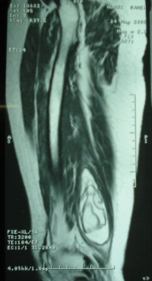
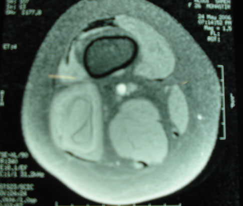
no history of abdominal or chest pain, coughing, hemopt- A wide excision of the cyst with a safety margin was ysis, fever, weight loss, and urticaria. The deep palpation of performed. No recurrence has been detected after a 3-year the posterior part of the right thigh revealed a soft tissue follow-up period. The authors review the literature and firm mass of 10 9 7 cm. There was no lymphadenopathy, discuss the MRI findings. The diagnosis of muscular and the local skin temperature was normal. The MRI hydatid cyst is done using clinical and radiological findings showed an 8 9 5 9 5 cm cystic lesion in the long head of in endemic context.
the biceps femoris muscle (Figs. , ). A thoraco-abdom-inopelvic computed tomography (CT) revealed no hydatid Hydatid cyst  Muscle  Surgery  MRI cyst involvement. Complete blood count was normal, butserology test (ELISA) for echinococcosis was positive(160 RU/ml).
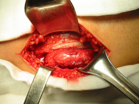
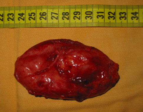
The treatment was surgical, under general anesthesia. The cyst was totally excised with a safety margin (Figs. ), and Echinococciasis or hydatid disease is a cosmopolitan followed by a 20% saline solution washing. The incision was anthropozoonosis common in man and several mammal primarily closed. Microscopic study confirmed the diagnosis species. It is endemic in most sheep-raising countries. The of hydatid disease. No medical treatment was given before liver and lungs are the most frequently affected organs.
and after surgery. After a 3-year follow-up, no recurrence Primary muscular hydatid cysts are extremely rare. We report a case of primary hydatid cyst of the long head of thebiceps femoris muscle with review of literature, and wediscuss the MRI findings.
Echinococciasis or hydatid disease is caused by larvalforms of small tapeworms of Echinococcus granulosis. Theincubation period may last 5–20 years, and symptoms mayshow up several years after exposure []. Larvae penetratethe intestinal mucosa and enter the portal blood stream;75% of cases are carried to the liver, 15% to the lungs, and M. F. Hamdi (&)  B. Touati  A. Abid the remainder to the rest of the body []. Musculoskeletal Department of Trauma and Orthopaedic Surgery, involvement accounts for only 1–4% of cases []; the F. Bourguiba University Hospital, 5000 Monastir, Tunisiae-mail: [email protected] muscle is considered an unfavorable site for hydatidosis


Musculoskelet Surg (2010) 94:59–61 Fig. 3 Operative view, the cyst was surrounded by a fibrous capsule(pericyst) Fig. 1 T1-weighted axial right thigh MRI showing low signalintensity; the signal intensity of the cyst is similar to that of muscles Fig. 4 Macroscopic appearance of the hydatid cyst after wideresection A preoperative radiological work-up may reveal the characteristic features of a hydatid cyst; radiological find-ings range from a purely cystic lesion to a completely solid Fig. 2 Coronal T2 MRI image of the right thigh with high signal appearance Magnetic resonance imaging is the diag- intensity of the cyst and water-lily sign; the low signal intensity nostic tool of choice. A continuous low-intensity rim with a surrounding the cyst representing the pericyst maximum thickness of 4–5 mm, multicystic appearance, ahomogeneous signal intensity of the cystic fluid and pres-ence of daughter cysts are the most characteristic MRI because of the contractility, the high lactic acid level and findings. The detachment of the germinative membrane the blood-filtering functions of the liver and lungs [, from pericyst (water-lily sign) is considered to be patho- The primary muscle hydatidosis without involvement of gnomonic Computerized tomography (CT) has been the thoracic and abdominal organs is extremely rare [, used in the diagnosis of subcutaneous hydatidosis, but MRI Given its relative rarity, the diagnosis of soft tissue hydatid is superior to CT in the final radiological diagnosis of cysts requires a high index of suspicion.
subcutaneous and muscular hydatidosis ]. Serological Musculoskelet Surg (2010) 94:59–61 tests and eosinophil count are not positive for echinococ- Echinococcosis endemic regions. The MRI characteristics cosis in all cases, they carry only an 80% of specificity, and may help in preoperative of muscular hydatid cyst diag- thus they may be helpful but not definitive in differential nosis. The diagnosis of muscular hydatid cyst is done diagnosis Several serological tests are available but using clinical and radiological findings in endemic may give negative findings because of encapsulation The treatment of muscle hydatid cyst is surgical using complete excision of the intact cyst. In fact cyst rupture can Conflict of interest cause anaphylactic shock and dissemination of the viablescolices to other organs and produce secondary cysts [, ]. Fine-needle aspiration biopsy of the hydatid cyst is a contemporary diagnostic procedure and may be used forpercutaneous aspiration and drainage. The complication of 1. Yu¨ksel BC, Akbulut S, Hengirmen S (2008) A minimally invasive this procedure (simple allergenic reactions to anaphylaxis) treatment option in primary muscular hydatid cyst: report of 2 usually occurs with non-intentional attempts at a rate of cases. Can J Surg 51(2):153–154 19% []. After excision of the cyst, the washing of the 2. Basarir K, Saglik Y, Yildiz Y, Yetis T, Cebesoy O (2008) Primary muscular hydatidosis mimicking soft tissue tumor: a report of five margin of the muscle with hypertonic saline solution was cases. J Orthop Surg (Hong Kong) 16(3):368–372 recommended to prevent the dissemination of the hydatid 3. Duygulu F, Karaog˘lu S, Erdog˘an N, Yildiz O (2006) Primary hydatid cyst of the thigh: a case report of an unusual localization.
Adjuvant administration of Albendazole (10 mg/kg twice Turk J Pediatr 48(3):256–259 4. Sarisoy HT, Memisoglu K, Tamer GS, Sarlak AY (2008) Primary daily) 3 or 2 months after surgery was uneventful to prevent hydatid disease in adductor muscles. Clin Invest Med 31(5):E296– local or distant dissemination [, but medical treatment alone is not effective []. The treatment of soft tissue hydatid 5. Erler K, Komurcu M, Ozmedir MT, Ozkal UB, Basbozkurt M, disease with Albendazole did not reduce the size of the Cunhan O (2003) Echinococcus infestation of quadriceps femorismuscle misdiagnosed as a soft tissue tumor (A case report). Turk J lesion, and a surgical excision was still necessary [].
Med Sci 33:127–130 6. Garcı´a-Alvarez F, Torcal J, Salinas JC, Gu¨emes A, Navarro AC, Lozano R (1999) Primary hydatid disease in lumbar muscles. Acta Orthop Belg 65(4):521–524 7. Ates M, Karakaplan M (2007) Hydatid cyst in the biceps and Gluteus muscles: case report. Surg Infect 8(4):475–478 Primary muscular hydatid cyst is extremely rare and 8. Ozkoc¸ G, Akpinar S, Hersekli MA, Ozalay M, Tandog˘an R (2003) should be included in the differential diagnosis of a slow- Primary hydatid disease of the quadriceps muscle: a rare locali- growing soft tissue mass of the extremities especially in zation. Arch Orthop Trauma Surg 123(6):314–316

Source: http://www.stcm.org.tn/upload/70.pdf

ppgia.pucpr.br

AgentSheets®: an Interactive Simulation Environment with End-User Programmable Agents Alexander Repenning AgentSheets Inc., Gunpark Drive 6560, Boulder, Colorado, 80301, email: [email protected] Center for LifeLong Learning and Design, Department of Computer Science, University of Colorado at Boulder, Campus Box 430, Boulder, Colorado 80309-0430, email: [email protected]

Ανασκόπηση

Review Ocular side-effects of urological Nikolaos A. Kostakopoulos, Vasileios G. Argyropoulos Department of Urology, IASO General Hospital, Athens, Greece Corresponding author: Nikolaos A. Kostakopoulos Email: '[email protected]' Η αυξανόμενη γήρανση του πληθυσμού έχει σαν αποτέλεσμα συχνότερη εμφάνιση συμπτωμάτων καλοήθους υπερπλασίας του προστάτη, ασταθούς κύστεως και στυτικής δυσλειτουργίας. Αυτό έχει σαν επακόλουθο ευρεία συνταγογράφηση. φαρμάκων, όπως αναστολέων της φωσφοδιεστεράσης, αντιμουσκαρινικών παραγόντων και α-αναστολέων που προκαλούν μερικές φορές σοβαρές παρενέργειες από τους οφθαλμούς. Σ΄αυτή την ανασκόπηση θα αναφερθούν περιληπτικά οι διάφορες δυνητικές οφθαλμικές παρενέργειες, η συχνότητά τους, η φυσική τους ιστορία και η σημασία τους για τον κλινικό γιατρό.