Fape.org.ph
The Commission on Higher Education
in collaboration with the Philippine Normal University
INITIAL RELEASE: 13 JUNE 2016
Teaching Guide for Senior High School GENERAL
SPECIALIZED SUBJECT ACADEMIC - STEM
This Teaching Guide was collaboratively developed and reviewed by
educators from public and private schools, colleges, and universities.
We encourage teachers and other education stakeholders to email their
feedback, comments, and recommendations to the Commission on
Higher Education, K to 12 Transition Program Management Unit -
Senior High School Support Team at [email protected]. We value your
feedback and recommendations.

Development Team
Team Leader: Florencia G. Claveria, Ph.D.,
Dawn T. Crisologo
Writers: Doreen D. Domingo, Ph.D., Janet S.
This Teaching Guide by the
Estacion, Ph.D., Mary Jane C. Flores, Ph.D.,
Commission on Higher Education is
licensed under a Creative Commons
Aileen C. dela Cruz, Chuckie Fer Calsado,
Nolasco H. Sablan, Justin Ray M. Guce
ShareAlike 4.0 International
Technical Editor: John Donnie A. Ramos, Ph.D.
License. This means you are free to:
Published by the Commission on Higher Education, 2016
Copy Reader: Joy R. Jimena
Share — copy and redistribute the
Chairperson: Patricia B. Licuanan, Ph.D.
material in any medium or format
Illustrators: Renan U. Ortiz, Daniela Louise B. Go
Adapt — remix, transform, and
Commission on Higher Education
Cover Artists: Paolo Kurtis N. Tan, Renan U. Ortiz
build upon the material.
K to 12 Transition Program Management Unit
The licensor, CHED, cannot revoke
Office Address: 4th Floor, Commission on Higher Education,
Senior High School Support Team
these freedoms as long as you
C.P. Garcia Ave., Diliman, Quezon City
CHED K to 12 Transition Program Management Unit
follow the license terms. However,
Telefax: (02) 441-0927 / E-mail Address: [email protected]
under the following terms:
Program Director: Karol Mark R. Yee
Attribution — You must give
Lead for Senior High School Support:
appropriate credit, provide a link to
the license, and indicate if changes
Gerson M. Abesamis
Consultants
were made. You may do so in any
Course Development Officers:
THIS PROJECT WAS DEVELOPED WITH THE PHILIPPINE NORMAL UNIVERSITY.
reasonable manner, but not in any
John Carlo P. Fernando, Danie Son D. Gonzalvo
way that suggests the licensor
University President: Ester B. Ogena, Ph.D.
endorses you or your use.
VP for Academics: Ma. Antoinette C. Montealegre, Ph.D.
Lead for Policy Advocacy and Communications:
NonCommercial — You may not use
VP for University Relations & Advancement: Rosemarievic V. Diaz, Ph.D.
Averill M. Pizarro
the material for commercial
Ma. Cynthia Rose B. Bautista, Ph.D., CHED
Teacher Training Officers:
ShareAlike — If you remix,
Bienvenido F. Nebres, S.J., Ph.D., Ateneo de Manila University
Ma. Theresa C. Carlos, Mylene E. Dones
transform, or build upon the
Carmela C. Oracion, Ph.D., Ateneo de Manila University
Monitoring and Evaluation Officer:
material, you must distribute your
Minella C. Alarcon, Ph.D., CHED
Robert Adrian N. Daulat
contributions under the same license
Gareth Price, Sheffield Hallam University
as the original.
Administrative Officers:
Stuart Bevins, Ph.D., Sheffield Hallam University
Ma. Leana Paula B. Bato, Kevin Ross D. Nera, Allison A. Danao, Ayhen Loisse B. Dalena Printed in the Philippines by EC-TEC Commercial, No. 32 St. Louis Compound 7, Baesa, Quezon City, [email protected]
Table of Contents
DepEd General Biology 1 Curriculum Guide . . . . . . . 5
Chapter 3: Energy Transformation
Lesson 11: Photosynthesis and Cellular Respiration . . . . . .
Lesson 1: The Cell: Endomembrane System, Mitochondria,
Lesson 12: Forms of Energy, Laws of Energy Transformation
Chloroplasts, Cytoskeleton, and Extracellular Components . . 9
Lesson 2: Mitochondria and Chloroplasts . . . . . . . . . 15
Lesson 13: Energy Transformation Part 1 . . . . . . . . . . 111
Lesson 3: Structure and Functions of Animal Tissues and Cell
Lesson 14: Energy Transformation Part 2 . . . . . . . . . . 120
Lesson 15: Energy Transformation Part 3 . . . . . . . . . . 128
Lesson 4: Cell Cycle and Cell Division . . . . . . . . . . 36
Lesson 16: Cellular Respiration Part 1 . . . . . . . . . . . 133
Lesson 5: Transport Mechanisms Part 1 . . . . . . . . . . 46
Lesson 17: Cellular Respiration Part 2 . . . . . . . . . . . 150
Lesson 6: Transport Mechanisms Part 2 . . . . . . . . . . 50
Lesson 18: Cellular Respiration Part 3 . . . . . . . . . . . 165
Chapter 2: Biological Molecules
Lesson 19: ATP in Cellular Metabolism and Photosynthesis . . . 176
Lesson 7: Carbohydrates and Lipids . . . . . . . . . . . 57
Lesson 8: Amino Acids and Proteins Part 1 . . . . . . . . 70
Lesson 9: Amino Acids and Proteins Part 2 . . . . . . . . 73
Lesson 10: Biological Molecules: Enzymes . . . . . . . . 78
As the Commission supports DepEd's implementation of Senior High School (SHS), it upholds the vision
and mission of the K to 12 program, stated in Section 2 of Republic Act 10533, or the Enhanced Basic
Education Act of 2013, that "every graduate of basic education be an empowered individual, through a
program rooted on.the competence to engage in work and be productive, the ability to coexist in
fruitful harmony with local and global communities, the capability to engage in creative and critical
thinking, and the capacity and willingness to transform others and oneself." To accomplish this, the Commission partnered with the Philippine Normal University (PNU), the National
Center for Teacher Education, to develop Teaching Guides for Courses of SHS. Together with PNU, this
Teaching Guide was studied and reviewed by education and pedagogy experts, and was enhanced with
appropriate methodologies and strategies. Furthermore, the Commission believes that teachers are the most important partners in attaining this
goal. Incorporated in this Teaching Guide is a framework that will guide them in creating lessons and
assessment tools, support them in facilitating activities and questions, and assist them towards deeper
content areas and competencies. Thus, the introduction of the SHS for SHS Framework.
The SHS for SHS Framework, which stands for "Saysay-Husay-Sarili for Senior High School," is at the
core of this book. The lessons, which combine high-quality content with flexible elements to
SHS for SHS accommodate diversity of teachers and environments, promote these three fundamental concepts:
SAYSAY: MEANING
HUSAY: MASTERY
SARILI: OWNERSHIP
Why is this important?
How will I deeply understand this?
What can I do with this?
Through this Teaching Guide,
Given that developing mastery
When teachers empower
teachers will be able to facilitate goes beyond memorization,
learners to take ownership of
an understanding of the value
teachers should also aim for
their learning, they develop
of the lessons, for each learner
deep understanding of the
independence and self-
to fully engage in the content
subject matter where they lead
direction, learning about both
on both the cognitive and
learners to analyze and
the subject matter and
affective levels.
synthesize knowledge.
Biology I is a Science, Technology, Engineering and Mathematics (STEM) Specialized Subject
taken in the first half of Grades 11/12. Learners go on a journey geared toward the deeper
understanding and appreciation of life processes at the cellular and molecular levels
previously introduced in Grades 7-10. They will also apply basic chemistry and physics
principles as they examine the transformation of energy in organisms. Implementing this course at the senior high school level is subject to numerous challenges
with mastery of content among educators tapped to facilitate learning and a lack of
resources to deliver the necessary content and develop skills and attitudes in the learners,
being foremost among these. In support of the SHS for SHS framework developed by CHED, these teaching guides were
crafted and refined by biologists and biology educators in partnership with educators from
focus groups all over the Philippines to provide opportunities to develop the following: Saysay through meaningful, updated, and context-specific content that highlights important
points and common misconceptions so that learners can connect to their real-world
experiences and future careers; Husay through diverse learning experiences that can be implemented in a resource-poor
classroom or makeshift laboratory that tap cognitive, affective, and psychomotor domains
are accompanied by field-tested teaching tips that aid in facilitating discovery and
development of higher-order thinking skills; and Sarili through flexible and relevant content and performance standards allow learners the
freedom to innovate, make their own decisions, and initiate activities to fully develop their
academic and personal potential. These ready-to-use guides are helpful to educators new to either the content or biologists
new to the experience of teaching Senior High School due to their enriched content
presented as lesson plans or guides. Veteran educators may also add ideas from these
guides to their repertoire. The Biology Team hopes that this resource may aid in easing the
transition of the different stakeholders into the new curriculum as we move towards the
constant improvement of Philippine education.
This Teaching Guide is mapped and aligned to the DepEd SHS Curriculum, designed to be highly
Parts of the
usable for teachers. It contains classroom activities and pedagogical notes, and is integrated with
innovative pedagogies. All of these elements are presented in the following parts:
Teaching Guide 1. Introduction
• Highlight key concepts and identify the essential questions
• Show the big picture
• Connect and/or review prerequisite knowledge
• Clearly communicate learning competencies and objectives
• Motivate through applications and connections to real-life
• Give local examples and applications
• Engage in a game or movement activity
• Provide a hands-on/laboratory activity
• Connect to a real-life problem
3. Instruction/Delivery
• Give a demonstration/lecture/simulation/hands-on activity
• Show step-by-step solutions to sample problems
• Give applications of the theory
• Connect to a real-life problem if applicable
• Discuss worked-out examples
• Provide easy-medium-hard questions
• Give time for hands-on unguided classroom work and discovery
• Use formative assessment to give feedback
• Provide additional examples and applications
• Introduce extensions or generalisations of concepts
• Engage in reflection questions
• Encourage analysis through higher order thinking prompts
• Supply a diverse question bank for written work and exercises
• Provide alternative formats for student work: written homework, journal, portfolio, group/individual
projects, student-directed research project
On DepEd Functional Skills and CHED College Readiness Standards
As Higher Education Institutions (HEIs) welcome the graduates of
On the other hand, the Commission declared the College
the Senior High School program, it is of paramount importance to
Readiness Standards that consist of the combination of knowledge,
align Functional Skills set by DepEd with the College Readiness
skills, and reflective thinking necessary to participate and succeed -
Standards stated by CHED.
without remediation - in entry-level undergraduate courses in
The DepEd articulated a set of 21st century skills that should be
embedded in the SHS curriculum across various subjects and tracks.
The alignment of both standards, shown below, is also presented in
These skills are desired outcomes that K to 12 graduates should
this Teaching Guide - prepares Senior High School graduates to the
possess in order to proceed to either higher education,
revised college curriculum which will initially be implemented by AY
employment, entrepreneurship, or middle-level skills development.
College Readiness Standards Foundational Skills
DepEd Functional Skills
Produce all forms of texts (written, oral, visual, digital) based on: 1. Solid grounding on Philippine experience and culture;
2. An understanding of the self, community, and nation;
Visual and information literacies, media literacy, critical thinking
3. Application of critical and creative thinking and doing processes;
and problem solving skills, creativity, initiative and self-direction
4. Competency in formulating ideas/arguments logically, scientifically, and creatively; and
5. Clear appreciation of one's responsibility as a citizen of a multicultural Philippines and a
Systematically apply knowledge, understanding, theory, and skills for the development of
Global awareness, scientific and economic literacy, curiosity,
the self, local, and global communities using prior learning, inquiry, and experimentation
critical thinking and problem solving skills, risk taking, flexibility
and adaptability, initiative and self-direction
Work comfortably with relevant technologies and develop adaptations and innovations for Global awareness, media literacy, technological literacy, significant use in local and global communities
creativity, flexibility and adaptability, productivity and
Communicate with local and global communities with proficiency, orally, in writing, and
Global awareness, multicultural literacy, collaboration and
through new technologies of communication
interpersonal skills, social and cross-cultural skills, leadership
and responsibility
Interact meaningfully in a social setting and contribute to the fulfilment of individual and
Media literacy, multicultural literacy, global awareness,
shared goals, respecting the fundamental humanity of all persons and the diversity of
collaboration and interpersonal skills, social and cross-cultural
skills, leadership and responsibility, ethical, moral, and spiritual
groups and communities
General Biology 1
The Cell: Endomembrane System, Mitochondria,
Chloroplasts, Cytoskeleton, and Extracellular Components
Content Standards
The learners demonstrate an understanding of (1) Composition of the
endomembrane system; (2) Structure and function of organelles involved in
Introduction Review on the differences between
energy transformation; (3) Structure and functions of the cytoskeleton; and, (4)
prokaryotic and eukaryotic cells; submission
Composition and functions of the extracellular components or matrix.
and discussion of responses to the pre-topic homework assigned before the lecture.
Performance Standards
The learners shall be able to construct three-dimensional models of whole cells Motivation
Brief class activity on prokaryotic and
using indigenous or recyclable materials. The models shall show the following
eukaryotic cells.
cell parts: (1) Endomembrane System, (2) Mitochondria, and (3) Chloroplast
Instruction/ Lecture. Board work on cell parts, structure,
Learning Competencies
and function. Examination of cheek cells and
The learners: (1) explain the postulates of the cell theory (STEM_BIO11/12-1a-
Hydrilla cells under a microscope. Class
c-1); (2) describe the structure and function of major and subcellular organelles
activity on identifying the parts and functions
(STEM_BIO11/12-Ia-c-2); (3) describe the structural components of the cell
of the endomembrane system.
membrane (STEM_BIO11/12-Ig-h-11); and (4) relate the structure and
Enrichment Class discussion on cell size and relationship
composition of the cell membrane to its function (STEM_BIO11/12-Ig-h-12)
of surface area and volume
Specific Learning Outcomes
Assessment of learners' knowledge;
At the end of the unit lesson, the learners shall be able to:
assignment of homework for next lecture
• illustrate the structure of the endomembrane system, label its parts, and
microscope (slide, cover slip), hand-held
understand how the system works
lens, work books, methylene blue, plastic
• illustrate the structure of the mitochondria, label its parts, and understand
spoon/popsicle stick, Hydrilla plansts,
the importance of the enfolding of the inner mitochondrial membrane
colored chalk/white board marker
• illustrate the structure of the chloroplast, label its parts, and relate these
parts to photosynthesis
Resources (continued at the end of Teaching Guide)
(1) (n.d.). Retrieved from <http://www.phschool.com/science/
• understand the connection of the endomembrane system to other cell
parts such as the lysosomes, peroxisomes, endosomes, and cell membrane (2) (n.d.). Retrieved from <http://biology.tutorvista.com/animal-and-plant-
• understand how the extracellular components or matrix determine the
(3) (n.d.). Retrieved from <http://sciencenetlinks.com/lessons/cells-2-the-
appearance and function of the tissues
INTRODUCTION (5 MINS)
Teacher Tip
The review on the differences between
1. Ask the learners to make a recap of the differences between prokaryotic and eukaryotic cells.
prokaryotic and eukaryotic cells is needed
2. Discuss the learners' responses to the pre-topic assignment on the functions of the following cell
to connect prerequisite knowledge to the
present lesson. Remind the learners that the
cell parts are found in eukaryotic cells.
• Nucleus • Smooth Endoplasmic Reticulum
Remind the learners of the pre-topic
assignment that shall be submitted before
• Rough Endoplasmic Reticulum
the lecture. This is to ensure the learners
• Golgi Apparatus
read on the topic before the lecture.
Briefly discuss the structure of the cell
membrane in order to provide basic
knowledge on said structure to the learners.
• Mitochondria
Do not fully elaborate on this topic since the
structure and function of the cell membrane
shall further be discussed in the succeeding
parts of the lesson.
3. Present an overview of the cell membrane, its structure, and functions.
The cell's parts should be discussed as a
4. Define what an ‘organelle' is and differentiate membrane-bound organelles from non-membrane-
system, emphasizing on the
bound organelles.
interconnectedness of each part to the
5. Explain that in eukaryotic cells, the machinery of the cell is compartmentalized into organelles. The
compartmentalization of the cell into membrane-bound organelles:
To clarify common misconceptions,
• allows conflicting functions (i.e., synthesis vs. breakdown) and several cellular activities to occur
emphasize the following to the learners:
simultaneously without interference from each other
• Not all organelles are surrounded by a
• separates the DNA material of the nucleus, mitochondria, and chloroplast
• The plasma or cell membrane is different
• increases the surface area-volume ratio of the cell
from the cell wall.
6. Encourage the learners to look at the cell as both a system and subsystem. They should develop an
• Not all cell parts are present in all kinds of
understanding of how the parts of a cell interact with one another and how these parts help to do the
‘work' of the cell (Source: (n.d.). Retrieved from <http://sciencenetlinks.com/lessons/cells-2-the-cell-as-
MOTIVATION (5 MINS)
Teacher tip
Briefly review the differences between prokaryotic and eukaryotic cells by asking questions to the
If the number of available microscopes is
limited, ask the learners to group
Sample question: What cell parts can be found in both prokaryotic and eukaryotic cells? Discuss themselves according to the number of
the function/s of each part.
microscopes available or set-up a
Sample Responses:
demonstration scope for the whole class
and facilitate the examination of cells so
that all the learners will get a chance to
observe the cells under the microscope.
• Cell membrane • Protoplasm (nucleoloid region and cytosol)
Orient the learners on the proper use and
care of the microscopes, particularly on
focusing first on LPO before shifting to
Compare the cell to a big city. Ask the learners what the requirements of the city would be in order for
Cheek cells are very transparent. Adjust the
it to function. Relate these requirements to the parts of the cell. Relate the learners' responses to the
iris diaphragm or add a small amount of dye
functions of the different parts of a cell.
(i.e., methylene blue) to the scrapings.
Sample responses:
The learners will only see the cell membrane
• The city will need power. What generates power for the city? Relate this to the function of
and the nucleus. Remind the learners to
the mitochondria and the chloroplast.
draw what they observe. Students may
observe cytoplasmic streaming in the plant
• The city generates waste. How does it minimize its waste? How does the city handle its
garbage? Relate this to the function of the lysosome.
• The city requires raw materials to process into food, clothing, and housing materials. Where
are these raw materials processed? Relate this to the functions of the Golgi Apparatus.
Compare animal cells from plant cells. For the animal cells, scrape cheek cells using a toothpick. Ask
the learners to place the scrapings on a microscope slide and add a drop of water to the scrapings.
Tease the scrapings into a thin layer and cover with a slip. Examine under HPO. Instruct the learners to
draw the cells on their workbooks and to label the cell parts that they were able to observe under the
microscope. For the plant cells, instruct the learners to obtain a Hydrilla leaf and place it on a microscope slide.
Examine under LPO. Ask the learners to draw the cells on their workbooks and to label the cell parts
that they were able to observe under the microscope.
INSTRUCTION/PRACTICE (30 MINS)
Teacher tip
Use chalk or white board markers with
different colors. Explain the structure and
1. Draw the cell membrane on one end of the board.
function of each cell part as you draw them.
2. Draw the double membrane of the nucleus (nuclear membrane) on the other end of the board.
Explain to the learners that a more detailed
3. From the nuclear membrane, draw the reticulated structure of the endoplasmic reticulum. Ask the
discussion of the structure and functions of
learners what the two types of endoplasmic reticulum are and their corresponding functions.
the cell membrane, mitochondria, and
4. Draw the ribosomes as separate units.
chloroplast will be given in succeeding
5. Draw a DNA and an mRNA. Explain that the mRNA is a copy of the DNA that will be sent to the
cytoplasm for protein synthesis.
6. Explain to the learners that the mRNA leaves the nucleus and goes to where the ribosomes are
located (i.e., mRNA + functional ribosome)
7. Explain the possible ‘pathways' for protein synthesis (e.g., within the cytosol or the endoplasmic
8. Draw the mRNA + functional ribosome on the endoplasmic reticulum. With a lot of these, the
endoplasmic reticulum becomes a rough endoplasmic reticulum.
9. Draw the formed polypeptide inside the rough endoplasmic reticulum. Discuss the formation of a
cisternae and pinching off as a vesicle.
10. Draw the Golgi Apparatus and then a vesicle from the rough endoplasmic reticulum that travels to
the Golgi Apparatus and attaches to the part which is nearest the rough endoplasmic reticulum.
11. Ask the learners what the function of the Golgi Apparatus is. Synthesize their answers and compare
the Golgi Apparatus to a factory with an assembly manufacturing line.
12. Draw the polypeptide travelling along the Golgi Apparatus stack; pinching off as a vesicle to travel
to the next stack. Repeat the process while increasing the complexity of the polypeptide drawing.
13. On the last stack, explain the ‘pathways' that the vesicle may follow: become a lysosome through
fusion with an endosome (i.e., formed by endocytosis), or travel to the cell membrane, fuse with it,
and empty its contents.
14. Present the composition of the endomembrane system and discuss how these parts are connected
to each other by structure and by function.
15. Draw the mitochondria and label its parts. Explain the importance of the enfolding (cristae) in
increasing the surface area of the inner mitochondrial membrane. Further explain to the class that
enfolding is a common structural strategy to increase surface area. As an example, you may draw a
cross-sectional structure of the small intestine.
16. Draw the chloroplast and label its parts. Explain the function that each part performs in the process
of photosynthesis.
17. Discuss the similarities of the mitochondria and chloroplast (e.g., both are involved in energy
transformation, both have DNA, high surface area, and double membranes).income accounts and
lastly, expenses accounts.
Group the learners into pairs. Ask one to draw the endomembrane system as he/she explains it to
his/her partner. Reshuffle the groupings and repeat until all learners have performed the exercise.
ENRICHMENT (30 MINS)
Facilitate a class discussion on why cells are generally small in size. Explain the relationship between
surface area and volume.
EVALUATION (60 MINS
Ask questions to the learners. Sample questions can be found in the following electronic resources:
Teacher tip
• (n.d.). Retrieved from< http://www.proprofs.com/quiz-school/story.php?title=cell-structure-test >
Assignments should be handwritten.
• (n.d.). Retrieved from< http://study.com/academy/exam/topic/cell-biology.html>
This strategy is aimed at ensuring that the
Assign a research assignment on this question: How do environmental toxins like lead and mercury
learners have read the topic rather than just
affect the functions of the cell? The assignment shall be submitted one week after this lesson.
copying and printing from a source.
RESOURCES (CONTINUED):
(4) (n.d.). Retrieved from <http://www.schools.manatee.k12.fl.us/072JOCONNOR/celllessonplans/lesson_plan cell_structure_and_function.html> (5) (n.d.). Retrieved from <http://www.phschool.com/science/biology_place/biocoach/cells/endo.html> (6) (n.d.). Retrieved from <http://study.com/academy/lesson/the-endomembrane-system-functions-components.html> (7) (n.d.). Retrieved from <http://www.ncbi.nlm.nih.gov/books/NBK26907/> (8) (n.d.). Retrieved from <http://staff.um.edu.mt/acus1/01Compart.pdf>
Learning Competency Assessment Tool
The learners shall be able
Learner was able to
Learner was able to answer Learner was able to
(1) Learner was not
participation (during answer all the question/s
the main question without
answer the questions able to answer the
1. describe the structure and lecture)
without referring to his/
referring to his/her notes
but he/she referred
function of major and
but was not able to answer to his/her notes
(2) Learner read notes
subcellular organelles
follow-up question/s
of his/her classmate
Learner submitted an
Learner submitted a
Learner submitted a
(1) Learner did not
assignment beyond the
comprehensive and well-
well written report
submit an assignment
written assignment
but some responses
(2) Learner submitted
a partially-finished assignment
The learners shall be able
Learner was able to
Learner was able to answer Learner was able to
(1) Learner was not
participation (during concisely answer all the
the main question without
answer the questions able to answer the
2. describe the structural
referring to his/her notes
but he/she referred
components of the cell
but was not able to answer to his/her notes
(2) Learner read notes
follow-up question/s
of his/her classmate
Learner submitted
Learner submitted drawings Learner submitted
(1) Learner was not
drawings that were
that fulfilled the
drawings that were
Animal and Plant
beyond the requirements
requirements (complete
(2) Learner's drawings were haphazardly done
The learners shall be able
Learner obtained 90% to
Learner obtained 70% to
Learner obtained
Learner obtained less
100% correct answers in
89.99% correct answers in
that 50% correct
3. relate the structure and
correct answers in the answers in the
composition of the cell
membrane to its function
Learner submitted a
Learner submitted a
Learner submitted a
(1) Learner did not
research assignment
comprehensive and well-
well written report
submit an assignment
beyond the requirements
written research assignment but some responses
(2) Learner submitted
a partially-finished assignment
General Biology 1
Mitochondria and Chloroplasts
Content Standards
The learners demonstrate an understanding of the structure and function of the
mitochondria and chloroplasts, the organelles involved in energy
Introduction Review of relevant terminologies and
Performance Standards
Understanding of key concepts using real-life
The learners shall be able to construct three-dimensional models of whole cells
using indigenous or recyclable materials. These models should show the
Instruction/ Discussion and lecture proper
mitochondria and chloroplasts.
Learning Competencies
Drawing (with label) activity
The learners describe the structure and function of major and subcellular
organelles (STEM_BIO11/12-Ia-c-2) and distinguish prokaryotic and eukaryotic Enrichment Computation of surface area vs volume
cells according to their distinguishing features (STEM_BIO11/12 -Ia-c-3)
Answering practice questions and homework
Resources (continued at the end of Teaching Guide)
Specific Learning Outcomes
At the end of the lesson, the learners shall be able to:
• illustrate the structure of the mitochondria, label its parts, and understand
the importance of the enfolding of the inner mitochondrial membrane
• illustrate the structure of the chloroplast, label its parts, and relate these
parts to photosynthesis
INTRODUCTION (5 MINS)
Facilitate a review of the following concepts: • Differences between prokaryotic and eukaryotic cells
• Definition of an ‘organelle'
• Differences between membrane-bound organelles and non-membrane-bound organelles
• Functions of the different parts of a cell
• The endomembrane system
Vacuoles and Vesicles
Chloroplast and other plastids
Explain that in eukaryotic cells, the machinery of the cell is compartmentalized into organelles. The compartmentalization of the cell into
membrane-bound organelles: • allows conflicting functions (i.e., synthesis vs. breakdown) and several cellular activities to occur simultaneously without interference from
• separates the DNA material of the nucleus, mitochondria, and chloroplast
• increases the surface area-volume ratio of the cell
Encourage the learners to look at the cell as both a system and subsystem. They should develop an
understanding of how the parts of a cell interact with one another and how these parts help to do the
‘work' of the cell (Source: (n.d.). Retrieved from <http://sciencenetlinks.com/lessons/cells-2-the-cell-as-
a-system/>) Emphasize to the learners that energy transformation is one of the characteristics of life. This refers to
the ability to obtain and use energy. This characterizes the main function of the mitochondria and the
MOTIVATION (5 MINS)
Ask the learners how they understand the concept of compartmentalization. Relate the concept to how
the cell is compartmentalized into organelles. Compare compartmentalization to the division of a house into a receiving room or sala, kitchen, dining
room, comfort rooms, bedrooms, etc.
Teacher tip
Ask the learners why they think a house is divided into several rooms.
Explain to the learner that this is how the
cell is able to allow conflicting functions
A possible response is that partitioning of the house into different parts facilitates the simultaneous
(e.g., synthesis vs breakdown) and several
occurrence of several activities without interfering with one another. Also, materials needed for each
cellular activities to occur simultaneously
activity can be stored at their specific areas. For example, pots and pans are being stored in the kitchen without interference from each other.
and not in the bedroom. Beds and pillows are found in the bedroom and not in the toilet/bath.
Explain to the learners that the mitochondria and chloroplasts have a small amount of DNA. Although
most of the proteins of these organelles are imported from the cytosol and are thus programmed by
the nuclear DNA, their DNA programs the synthesis of the proteins made on the organelles' ribosomes
(Source: Campbell et al). Compartmentalization separates the DNA material of the nucleus,
mitochondria, and chloroplast. Ask the learners if they have experienced going to a city/municipal hall and if they have observed that
the Mayor, Vice-Mayor, and the City/Municipal Administrator have separate offices. You can use other
examples such as the University President, VP for Academic Affairs, VP for Finance; Philippine
President, Vice President, Senators, etc. Compare the nuclear DNA to the Mayor and the mitochondrial DNA and chloroplast DNA to the Vice
Mayor. The Mayor runs the city/municipality but the Vice Mayor also performs functions that are
Teacher tip
specific to their positions. They need different offices (or compartments) to avoid conflict in their
Select a fruit that can be easily peeled like
calamansi or dalandan
Introduce the concept of surface area-volume ratio/relationship to the learners. Show a fruit to the
learners and explain that the outer surface of the fruit is the surface area. Peel the fruit and show them
what's inside, explaining that the inside of the fruit is the volume.
Explain to the learners that surface area (SA) and volume (V) do not increase in the same manner. As an
object increases in size, its volume increases as the cube of its linear dimensions while surface area
increases as the square of its linear dimensions. Example: If the initial starting point is the same: SA = 2; Volume = 2 (Ratio = 1:1) A one-step increase will result to: SA = 22 = 4 while V = 23 = 8 (Ratio = 1:2)
Teacher tip
INSTRUCTION/DELIVERY (30 MINS)
Ask questions to the learners while giving
Explain and discuss the nature and functions of the Adenosine Triphosphate (ATP) to the learners.
If an LCD projector is not available, draw
the structure of the mitochondria and
Adenosine Triphosphate (ATP)—It is the major energy currency of the cell that provides the energy for chloroplast on the board.
most of the energy-consuming activities of the cell. The ATP regulates many biochemical pathways. Mechanism: When the third phosphate group of ATP is removed by hydrolysis, a substantial amount of
free energy is released. ATP + H2O → ADP + Pi where ADP is adenosine diphosphate and Pi is inorganic phosphate Group the learners into pairs. Ask one to draw the endomembrane system as he/she explains it to his/
her partner. Reshuffle the groupings and repeat until all learners have performed the exercise.
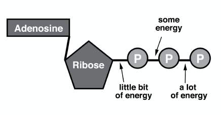
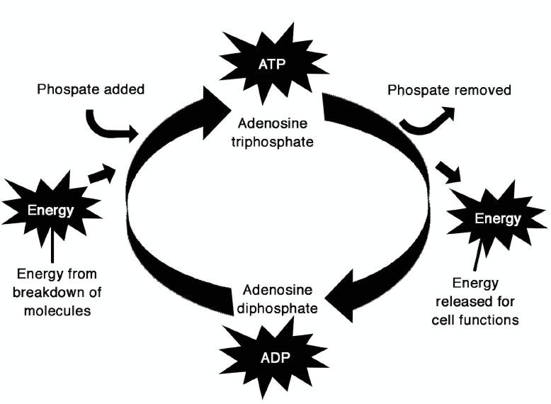
Illustration 1: Energy release in Hydrolysis (Source: (n.d.). Retrieved fr
Illustration 2: Chemical Energy and ATP (Source: (n.d.). Retrieved from
Synthesis of ATP
• ADP + Pi → ATP + H2O
• requires energy: 7.3 kcal/mole
• occurs in the cytosol by glycolysis
• occurs in mitochondria by cellular respiration
• occurs in chloroplasts by photosynthesis
Consumption of ATP
ATP powers most energy-consuming activities of cells, such as:
• anabolic (synthesis) reactions, such as:
• joining transfer RNAs to amino acids for assembly into proteins
• synthesis of nucleoside triphosphates for assembly into DNA and RNA
• synthesis of polysaccharides
• synthesis of fats
• active transport of molecules and ions
• conduction of nerve impulses
• maintenance of cell volume by osmosis • addition of phosphate groups (phosphorylation) to different proteins (e.g., to alter their activity in cell
• muscle contraction
• beating of cilia and flagella (including sperm)
• bioluminescence
Extracellular ATP In mammals, ATP also functions outside of cells. ATP is released in the following examples: • from damaged cells to elicit inflammation and pain
• from the carotid body to signal a shortage of oxygen in the blood
• from taste receptor cells to trigger action potentials in the sensory nerves leading back to the brain
• from the stretched wall of the urinary bladder to signal when the bladder needs emptying
In eukaryotic cells, the mitochondria and chloroplasts are the organelles that convert energy to other
forms which cells can use for their functions.
Discuss the function and structure of the mitochondria.
Mitochondria (singular, mitochondrion)—Mitochondria are the sites of cellular respiration, the
metabolic process that uses oxygen to drive the generation of ATP by extracting energy from sugars,
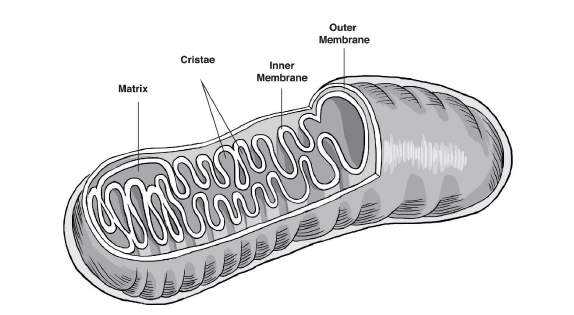
fats, and other fuels. The mitochondria are oval-shaped organelles found in most eukaryotic cells. They are considered to be
the ‘powerhouses' of the cell. As the site of cellular respiration, mitochondria serve to transform
molecules such as glucose into an energy molecule known as adenosine triphosphate (ATP). ATP fuels
cellular processes by breaking its high-energy chemical bonds. Mitochondria are most plentiful in cells
that require significant amounts of energy to function, such as liver and muscle cells. Figure 1: Structure of the Mitochonsdria (Source: (n.d.). Retrieved from http://www.britannica.com/list/
The mitochondria has two membranes that are similar in composition to the cell membrane: • Outer membrane—is a selectively permeable membrane that surrounds the mitochondria. It is the
site of attachment for the respiratory assembly of the electron transport chain and ATP Synthase. It
has integral proteins and pores for transporting molecules just like the cell membrane
• Inner membrane—folds inward (called cristae) to increase surfaces for cellular metabolism. It
contains ribosomes and the DNA of the mitochondria. The inner membrane creates two enclosed
spaces within the mitochondria: • intermembrane space between the outer membrane and the inner membrane; and
• matrix that is enclosed within the inner membrane.
Ask questions to the learners on the structure of the mitochondria. A sample question could be: What
is the importance of the enfolding of the mitochondria? The response would be to increase the surface
area that can be ‘packed' into such a small space.
Discuss the purpose of the mitochondrial membranes.
As mentioned, the mitochondria has two membranes: the outer and inner mitochondrial membranes. • Outer Membrane
Teacher tip
• fully surrounds the inner membrane, with a small intermembrane space in between
• has many protein-based pores that are big enough to allow the passage of ions and
molecules as large as a small protein
Lecture on mitochondrial membranes can
• Inner membrane
be accessed at (n.d.). Retrieved from
• has restricted permeability like the plasma membrane
• is loaded with proteins involved in electron transport and ATP synthesis
• surrounds the mitochondrial matrix, where the citric acid cycle produces the electrons that
travel from one protein complex to the next in the inner membrane. At the end of this
electron transport chain, the final electron acceptor is oxygen, and this ultimately forms
water (H20). At the same time, the electron transport chain produces ATP in a process called
oxidative phosphorylation
During electron transport, the participating protein complexes push protons from the matrix out to the
intermembrane space. This creates a concentration gradient of protons that another protein complex,
called ATP synthase, uses to power synthesis of the energy carrier molecule ATP. Figure 4: The Electrochemical Proton Gradient and the ATP Synthase (Source: (n.d.). Retrieved from
Explain and discuss the structure and functions of the Chloroplasts. Chloroplasts—Chloroplasts, which are found in plants and algae, are the sites of photosynthesis. This
process converts solar energy to chemical energy by absorbing sunlight and using it to drive the
synthesis of organic compounds such as sugars from carbon dioxide and water.
The word chloroplast is derived from the Greek word chloros which means ‘green' and plastes which
means ‘the one who forms'. The chloroplasts are cellular organelles of green plants and some
eukaryotic organisms. These organelles conduct photosynthesis. They absorb sunlight and convert it
into sugar molecules. They also produce free energy stored in the form of ATP and NADPH through
photosynthesis. Chloroplasts are double membrane-bound organelles and are the sites of photosynthesis. The
chloroplast has a system of three membranes: the outer membrane, the inner membrane, and the
thylakoid system. The outer and the inner membranes of the chloroplast enclose a semi-gel-like fluid
Teacher tip
known as the stroma. The stroma makes up much of the volume of the chloroplast. The thylakoid
system floats in the stroma.
If an LCD projector is not available, draw
the structure of the chloroplast on the
Structure of the Chloroplast
• Outer membrane—This is a semi-porous membrane and is permeable to small molecules and ions
which diffuse easily. The outer membrane is not permeable to larger proteins.
• Intermembrane Space—This is usually a thin intermembrane space about 10-20 nanometers and is
present between the outer and the inner membrane of the chloroplast.
• Inner membrane—The inner membrane of the chloroplast forms a border to the stroma. It
Lecture on structure and functions of the
regulates passage of materials in and out of the chloroplast. In addition to the regulation activity,
chloroplast can be accessed at (n.d.).
fatty acids, lipids and carotenoids are synthesized in the inner chloroplast membrane.
Retrieved from <http://
• Stroma—This is an alkaline, aqueous fluid that is protein-rich and is present within the inner
membrane of the chloroplast. It is the space outside the thylakoid space. The chloroplast DNA,
chloroplast ribosomes, thylakoid system, starch granules, and other proteins are found floating
around the stroma.
• Thylakoid System
The thylakoid system is suspended in the stroma. It is a collection of membranous sacks called
thylakoids. Thylakoids are small sacks that are interconnected. The membranes of these thylakoids are
the sites for the light reactions of the photosynthesis to take place. The chlorophyll is found in the
thylakoids. The thylakoids are arranged in stacks known as grana. Each granum contains around 10-20
thylakoids. The word thylakoid is derived from the Greek word thylakos which means 'sack'.
Important protein complexes which carry out the light reaction of photosynthesis are embedded in the
membranes of the thylakoids.
The Photosystem I and the Photosystem II are
complexes that harvest light with chlorophyll and carotenoids. They
absorb the light energy and use it to energize the electrons.
The molecules present in the thylakoid membrane use the electrons
that are energized to pump hydrogen ions into the thylakoid space.
This decreases the pH and causes it to become acidic in nature. A
large protein complex known as the ATP synthase controls the
concentration gradient of the hydrogen ions in the thylakoid space
to generate ATP energy. The hydrogen ions flow back into the
Thylakoids are of two types: granal thylakoids and stromal
PRACTICE (10 MINS)
thylakoids. Granal thylakoids are arranged in the grana. These
circular discs that are about 300-600 nanometers in diameter. The
Group the learners into pairs. Ask one to draw the mitochondria and
stromal thylakoids are in contact with the stroma and are in the form
label its parts while the other does the same for chloroplast. Once
of helicoid sheets.
done, the partners exchange tasks (i.e., the learner that drew the
mitochondria now does the same for the chloroplast).
The granal thylakoids contain only Photosystem II protein complex.
This allows them to stack tightly and form many granal layers with
Reproduce these diagrams without the labels and use these for the
granal membrane. This structure increases stability and surface area
for the capture of light.
To demonstrate how folding increases surface area, ask the learners
to trace the edges of the outer membrane with a thread and
The Photosystem I and ATP synthase protein complexes are present
measure the length of the thread afterwards. Repeat the same for
in the stroma. These protein complexes act as spacers between the
the inner membrane. Compare the results and discuss how the
sheets of stromal thylakoids.
enfolding of the inner membrane increases surface area through

ENRICHMENT (30 MINS)
1. Using the figure below, ask learners to compute surface area vs. volume. 2. Draw the table on the board and instruct the learners to write their measurements.
Teacher tip
EVALUATION (60 MINS)
Clarify to the learners the
Ask the learners to answer practice questions on the following electronic resources:
misconception that the appearance of
organelles are static and rigid.
Possible responses to the homework (Source: Campbell et al, 10th Ed.):
Teacher tip
Check the electronic resources on
• They have double membranes and are not part of the endomembrane system.
Endosymbiotic Theory:
• Their shape is changeable.
• They are autonomous (somewhat independent) organelles that grow and occasionally pinch in two,
thereby reproducing themselves.
v=bBjD4A7R2xU (Endosymbiotic
Theory in plain English)
• They are mobile and move around the cell along tracks of the cytoskeleton, a structural network of the
• They contain ribosomes, as well as multiple circular DNA molecules associated with their inner
membranes. The DNA in these organelles programs the synthesis of some organelle proteins on
ribosomes that have been synthesized and assembled there as well.
2. Give out the homework for next meeting.
What are the characteristics shared by these two energy transforming organelles?
Instruct the learners to write an essay on probable reasons for these the shared characteristics of the
mitochondria and the chloroplast. Learners shall submit a handwritten essay on the Endosymbiotic Theory
and how it explains the similarity between the mitochondria and chloroplast.
Learning Competency Assessment Tool
The learners shall be
Learner was able to
Learner was able to
Learner was able to (1) Learner was not
able to describe the
answer all the question/ answer the main question answer the
able to answer the
s without referring to
without referring to his/
questions but he/
her notes but was not
she referred to his/
(2) Learner read
able to answer follow-up her notes
1. structure and
notes of his/her
function of major and
subcellular organelles Assignment
Learner submitted an
Learner submitted a
Learner submitted a (1) Learner did not
assignment beyond the comprehensive and well- well written report
written assignment
but some responses assignment
partially-finished
Learner obtained 90%
Learner obtained 70% to Learner obtained
Learner obtained
89.99% correct answers
less that 50% correct
in the examination
correct answers in
Essay Assignment Learner submitted an
Learner submitted an
Learner submitted a (1) Learner did not
essay beyond the
well-written essay
comprehensive and well- some details are
partially-finished
General Biology 1
Mitochondria and Chloroplasts
Content Standards
The learners demonstrate an understanding of the structure and function of the
mitochondria and chloroplasts, the organelles involved in energy
Introduction Review of relevant terminologies and
Performance Standards
Understanding of key concepts using real-life
The learners shall be able to construct three-dimensional models of whole cells
using indigenous or recyclable materials. These models should show the
Instruction/ Discussion and lecture proper
mitochondria and chloroplasts.
Learning Competencies
Drawing (with label) activity
The learners describe the structure and function of major and subcellular
organelles (STEM_BIO11/12-Ia-c-2) and distinguish prokaryotic and eukaryotic Enrichment Computation of surface area vs volume
cells according to their distinguishing features (STEM_BIO11/12 -Ia-c-3)
Answering practice questions and homework
Resources (continued at the end of Teaching Guide)
Specific Learning Outcomes
At the end of the lesson, the learners shall be able to:
• illustrate the structure of the mitochondria, label its parts, and understand
the importance of the enfolding of the inner mitochondrial membrane
• illustrate the structure of the chloroplast, label its parts, and relate these
parts to photosynthesis
INTRODUCTION (5 MINS)
Facilitate a review of the following concepts: • Differences between prokaryotic and eukaryotic cells
• Definition of an ‘organelle'
• Differences between membrane-bound organelles and non-membrane-bound organelles
• Functions of the different parts of a cell
• The endomembrane system
Vacuoles and Vesicles
Chloroplast and other plastids
Explain that in eukaryotic cells, the machinery of the cell is compartmentalized into organelles. The compartmentalization of the cell into
membrane-bound organelles: • allows conflicting functions (i.e., synthesis vs. breakdown) and several cellular activities to occur simultaneously without interference from
• separates the DNA material of the nucleus, mitochondria, and chloroplast
• increases the surface area-volume ratio of the cell
Encourage the learners to look at the cell as both a system and subsystem. They should develop an
understanding of how the parts of a cell interact with one another and how these parts help to do the
‘work' of the cell (Source: (n.d.). Retrieved from <http://sciencenetlinks.com/lessons/cells-2-the-cell-as-
a-system/>) Emphasize to the learners that energy transformation is one of the characteristics of life. This refers to
the ability to obtain and use energy. This characterizes the main function of the mitochondria and the
MOTIVATION (5 MINS)
Ask the learners how they understand the concept of compartmentalization. Relate the concept to how
the cell is compartmentalized into organelles. Compare compartmentalization to the division of a house into a receiving room or sala, kitchen, dining
room, comfort rooms, bedrooms, etc.
Teacher tip
Ask the learners why they think a house is divided into several rooms.
Explain to the learner that this is how the
cell is able to allow conflicting functions
A possible response is that partitioning of the house into different parts facilitates the simultaneous
(e.g., synthesis vs breakdown) and several
occurrence of several activities without interfering with one another. Also, materials needed for each
cellular activities to occur simultaneously
activity can be stored at their specific areas. For example, pots and pans are being stored in the kitchen without interference from each other.
and not in the bedroom. Beds and pillows are found in the bedroom and not in the toilet/bath.
Explain to the learners that the mitochondria and chloroplasts have a small amount of DNA. Although
most of the proteins of these organelles are imported from the cytosol and are thus programmed by
the nuclear DNA, their DNA programs the synthesis of the proteins made on the organelles' ribosomes
(Source: Campbell et al). Compartmentalization separates the DNA material of the nucleus,
mitochondria, and chloroplast. Ask the learners if they have experienced going to a city/municipal hall and if they have observed that
the Mayor, Vice-Mayor, and the City/Municipal Administrator have separate offices. You can use other
examples such as the University President, VP for Academic Affairs, VP for Finance; Philippine
President, Vice President, Senators, etc. Compare the nuclear DNA to the Mayor and the mitochondrial DNA and chloroplast DNA to the Vice
Mayor. The Mayor runs the city/municipality but the Vice Mayor also performs functions that are
Teacher tip
specific to their positions. They need different offices (or compartments) to avoid conflict in their
Select a fruit that can be easily peeled like
calamansi or dalandan
Introduce the concept of surface area-volume ratio/relationship to the learners. Show a fruit to the
learners and explain that the outer surface of the fruit is the surface area. Peel the fruit and show them
what's inside, explaining that the inside of the fruit is the volume.
Explain to the learners that surface area (SA) and volume (V) do not increase in the same manner. As an
object increases in size, its volume increases as the cube of its linear dimensions while surface area
increases as the square of its linear dimensions. Example: If the initial starting point is the same: SA = 2; Volume = 2 (Ratio = 1:1) A one-step increase will result to: SA = 22 = 4 while V = 23 = 8 (Ratio = 1:2)
Teacher tip
INSTRUCTION/DELIVERY (30 MINS)
Ask questions to the learners while giving
Explain and discuss the nature and functions of the Adenosine Triphosphate (ATP) to the learners.
If an LCD projector is not available, draw
the structure of the mitochondria and
Adenosine Triphosphate (ATP)—It is the major energy currency of the cell that provides the energy for chloroplast on the board.
most of the energy-consuming activities of the cell. The ATP regulates many biochemical pathways. Mechanism: When the third phosphate group of ATP is removed by hydrolysis, a substantial amount of
free energy is released. ATP + H2O → ADP + Pi where ADP is adenosine diphosphate and Pi is inorganic phosphate Group the learners into pairs. Ask one to draw the endomembrane system as he/she explains it to his/
her partner. Reshuffle the groupings and repeat until all learners have performed the exercise.
Illustration 1: Energy release in Hydrolysis (Source: (n.d.). Retrieved fr
Illustration 2: Chemical Energy and ATP (Source: (n.d.). Retrieved from
Synthesis of ATP
• ADP + Pi → ATP + H2O
• requires energy: 7.3 kcal/mole
• occurs in the cytosol by glycolysis
• occurs in mitochondria by cellular respiration
• occurs in chloroplasts by photosynthesis
Consumption of ATP
ATP powers most energy-consuming activities of cells, such as:
• anabolic (synthesis) reactions, such as:
• joining transfer RNAs to amino acids for assembly into proteins
• synthesis of nucleoside triphosphates for assembly into DNA and RNA
• synthesis of polysaccharides
• synthesis of fats
• active transport of molecules and ions
• conduction of nerve impulses
• maintenance of cell volume by osmosis • addition of phosphate groups (phosphorylation) to different proteins (e.g., to alter their activity in cell
• muscle contraction
• beating of cilia and flagella (including sperm)
• bioluminescence
Extracellular ATP In mammals, ATP also functions outside of cells. ATP is released in the following examples: • from damaged cells to elicit inflammation and pain
• from the carotid body to signal a shortage of oxygen in the blood
• from taste receptor cells to trigger action potentials in the sensory nerves leading back to the brain
• from the stretched wall of the urinary bladder to signal when the bladder needs emptying
In eukaryotic cells, the mitochondria and chloroplasts are the organelles that convert energy to other
forms which cells can use for their functions.
Discuss the function and structure of the mitochondria.
Mitochondria (singular, mitochondrion)—Mitochondria are the sites of cellular respiration, the
metabolic process that uses oxygen to drive the generation of ATP by extracting energy from sugars,
fats, and other fuels. The mitochondria are oval-shaped organelles found in most eukaryotic cells. They are considered to be
the ‘powerhouses' of the cell. As the site of cellular respiration, mitochondria serve to transform
molecules such as glucose into an energy molecule known as adenosine triphosphate (ATP). ATP fuels
cellular processes by breaking its high-energy chemical bonds. Mitochondria are most plentiful in cells
that require significant amounts of energy to function, such as liver and muscle cells. Figure 1: Structure of the Mitochonsdria (Source: (n.d.). Retrieved from http://www.britannica.com/list/
The mitochondria has two membranes that are similar in composition to the cell membrane: • Outer membrane—is a selectively permeable membrane that surrounds the mitochondria. It is the
site of attachment for the respiratory assembly of the electron transport chain and ATP Synthase. It
has integral proteins and pores for transporting molecules just like the cell membrane
• Inner membrane—folds inward (called cristae) to increase surfaces for cellular metabolism. It
contains ribosomes and the DNA of the mitochondria. The inner membrane creates two enclosed
spaces within the mitochondria: • intermembrane space between the outer membrane and the inner membrane; and
• matrix that is enclosed within the inner membrane.
Ask questions to the learners on the structure of the mitochondria. A sample question could be: What
is the importance of the enfolding of the mitochondria? The response would be to increase the surface
area that can be ‘packed' into such a small space.
Discuss the purpose of the mitochondrial membranes.
As mentioned, the mitochondria has two membranes: the outer and inner mitochondrial membranes. • Outer Membrane
Teacher tip
• fully surrounds the inner membrane, with a small intermembrane space in between
• has many protein-based pores that are big enough to allow the passage of ions and
molecules as large as a small protein
Lecture on mitochondrial membranes can
• Inner membrane
be accessed at (n.d.). Retrieved from
• has restricted permeability like the plasma membrane
• is loaded with proteins involved in electron transport and ATP synthesis
• surrounds the mitochondrial matrix, where the citric acid cycle produces the electrons that
travel from one protein complex to the next in the inner membrane. At the end of this
electron transport chain, the final electron acceptor is oxygen, and this ultimately forms
water (H20). At the same time, the electron transport chain produces ATP in a process called
oxidative phosphorylation
During electron transport, the participating protein complexes push protons from the matrix out to the
intermembrane space. This creates a concentration gradient of protons that another protein complex,
called ATP synthase, uses to power synthesis of the energy carrier molecule ATP. Figure 4: The Electrochemical Proton Gradient and the ATP Synthase (Source: (n.d.). Retrieved from
Explain and discuss the structure and functions of the Chloroplasts. Chloroplasts—Chloroplasts, which are found in plants and algae, are the sites of photosynthesis. This
process converts solar energy to chemical energy by absorbing sunlight and using it to drive the
synthesis of organic compounds such as sugars from carbon dioxide and water.
The word chloroplast is derived from the Greek word chloros which means ‘green' and plastes which
means ‘the one who forms'. The chloroplasts are cellular organelles of green plants and some
eukaryotic organisms. These organelles conduct photosynthesis. They absorb sunlight and convert it
into sugar molecules. They also produce free energy stored in the form of ATP and NADPH through
photosynthesis. Chloroplasts are double membrane-bound organelles and are the sites of photosynthesis. The
chloroplast has a system of three membranes: the outer membrane, the inner membrane, and the
thylakoid system. The outer and the inner membranes of the chloroplast enclose a semi-gel-like fluid
Teacher tip
known as the stroma. The stroma makes up much of the volume of the chloroplast. The thylakoid
system floats in the stroma.
If an LCD projector is not available, draw
the structure of the chloroplast on the
Structure of the Chloroplast
• Outer membrane—This is a semi-porous membrane and is permeable to small molecules and ions
which diffuse easily. The outer membrane is not permeable to larger proteins.
• Intermembrane Space—This is usually a thin intermembrane space about 10-20 nanometers and is
present between the outer and the inner membrane of the chloroplast.
• Inner membrane—The inner membrane of the chloroplast forms a border to the stroma. It
Lecture on structure and functions of the
regulates passage of materials in and out of the chloroplast. In addition to the regulation activity,
chloroplast can be accessed at (n.d.).
fatty acids, lipids and carotenoids are synthesized in the inner chloroplast membrane.
Retrieved from <http://
• Stroma—This is an alkaline, aqueous fluid that is protein-rich and is present within the inner
membrane of the chloroplast. It is the space outside the thylakoid space. The chloroplast DNA,
chloroplast ribosomes, thylakoid system, starch granules, and other proteins are found floating
around the stroma.
• Thylakoid System
The thylakoid system is suspended in the stroma. It is a collection of membranous sacks called
thylakoids. Thylakoids are small sacks that are interconnected. The membranes of these thylakoids are
the sites for the light reactions of the photosynthesis to take place. The chlorophyll is found in the
thylakoids. The thylakoids are arranged in stacks known as grana. Each granum contains around 10-20
thylakoids. The word thylakoid is derived from the Greek word thylakos which means 'sack'.
Important protein complexes which carry out the light reaction of photosynthesis are embedded in the
membranes of the thylakoids.
The Photosystem I and the Photosystem II are
complexes that harvest light with chlorophyll and carotenoids. They
absorb the light energy and use it to energize the electrons.
The molecules present in the thylakoid membrane use the electrons
that are energized to pump hydrogen ions into the thylakoid space.
This decreases the pH and causes it to become acidic in nature. A
large protein complex known as the ATP synthase controls the
concentration gradient of the hydrogen ions in the thylakoid space
to generate ATP energy. The hydrogen ions flow back into the
Thylakoids are of two types: granal thylakoids and stromal
PRACTICE (10 MINS)
thylakoids. Granal thylakoids are arranged in the grana. These
circular discs that are about 300-600 nanometers in diameter. The
Group the learners into pairs. Ask one to draw the mitochondria and
stromal thylakoids are in contact with the stroma and are in the form
label its parts while the other does the same for chloroplast. Once
of helicoid sheets.
done, the partners exchange tasks (i.e., the learner that drew the
mitochondria now does the same for the chloroplast).
The granal thylakoids contain only Photosystem II protein complex.
This allows them to stack tightly and form many granal layers with
Reproduce these diagrams without the labels and use these for the
granal membrane. This structure increases stability and surface area
for the capture of light.
To demonstrate how folding increases surface area, ask the learners
to trace the edges of the outer membrane with a thread and
The Photosystem I and ATP synthase protein complexes are present
measure the length of the thread afterwards. Repeat the same for
in the stroma. These protein complexes act as spacers between the
the inner membrane. Compare the results and discuss how the
sheets of stromal thylakoids.
enfolding of the inner membrane increases surface area through
ENRICHMENT (30 MINS)
1. Using the figure below, ask learners to compute surface area vs. volume. 2. Draw the table on the board and instruct the learners to write their measurements.
Teacher tip
EVALUATION (60 MINS)
Clarify to the learners the
Ask the learners to answer practice questions on the following electronic resources:
misconception that the appearance of
organelles are static and rigid.
Possible responses to the homework (Source: Campbell et al, 10th Ed.):
Teacher tip
Check the electronic resources on
• They have double membranes and are not part of the endomembrane system.
Endosymbiotic Theory:
• Their shape is changeable.
• They are autonomous (somewhat independent) organelles that grow and occasionally pinch in two,
thereby reproducing themselves.
v=bBjD4A7R2xU (Endosymbiotic
Theory in plain English)
• They are mobile and move around the cell along tracks of the cytoskeleton, a structural network of the
• They contain ribosomes, as well as multiple circular DNA molecules associated with their inner
membranes. The DNA in these organelles programs the synthesis of some organelle proteins on
ribosomes that have been synthesized and assembled there as well.
2. Give out the homework for next meeting.
What are the characteristics shared by these two energy transforming organelles?
Instruct the learners to write an essay on probable reasons for these the shared characteristics of the
mitochondria and the chloroplast. Learners shall submit a handwritten essay on the Endosymbiotic Theory
and how it explains the similarity between the mitochondria and chloroplast.
Learning Competency Assessment Tool
The learners shall be
Learner was able to
Learner was able to
Learner was able to (1) Learner was not
able to describe the
answer all the question/ answer the main question answer the
able to answer the
s without referring to
without referring to his/
questions but he/
her notes but was not
she referred to his/
(2) Learner read
able to answer follow-up her notes
1. structure and
notes of his/her
function of major and
subcellular organelles Assignment
Learner submitted an
Learner submitted a
Learner submitted a (1) Learner did not
assignment beyond the comprehensive and well- well written report
written assignment
but some responses assignment
partially-finished
Learner obtained 90%
Learner obtained 70% to Learner obtained
Learner obtained
89.99% correct answers
less that 50% correct
in the examination
correct answers in
Essay Assignment Learner submitted an
Learner submitted an
Learner submitted a (1) Learner did not
essay beyond the
well-written essay
comprehensive and well- some details are
partially-finished
General Biology 1
Structure and Functions of Animal Tissues
and Cell Modification
Content Standard
The learners demonstrate an understanding of animal tissues and cell
Introduction Communicating learning objectives to the
Performance Standard
Class Activity: Pinoy Henyo Classroom
The learners shall be able to construct a three-dimensional model of the animal
tissue by using recyclable or indigenous materials.
Instruction/ Review on the Hierarchy of Biological
Learning Competencies
Organisation and PTSF; Lesson on Animal
Tissues and on Cell Modfication
• classify different cell types (plant/animal tissue) and specify the functions of
Class Activity: Reporting on structure and
each (STEM_BIO11/12-Ia-c-4)
function of animal tissue or showing of
• describe some cell modifications that lead to adaptation to carry out
infomercial on diseases.
specialized functions (e.g., microvilli, root hair) (STEM_BIO11/12-Ia-c-5)
Specific Learning Outcomes
At the end of the lesson, the learners shall be able to:
Materials
• present a five-minute report on how the structures of different animal
microscopes, LCD Projector (if available), laptop or computer
tissues define their function or show a two-minute infomercial about a
(if available), manila paper, cartolina, photos, images, or
disease that is caused by animal tissue malfunction;
illustrations of different types of tissues, drawing materials
• provide insights, offer constructive feedback, and note areas of
(e.g. pens, pencils, paper, color pencils, etc.)
improvement on their classmates' reports or infomercial
Resources (continued at the end of Teaching Guide)
(1) Reece JB, U. L., (2010). Campbell Biology 10th. San Francisco (CA).
INTRODUCTION (5 MINS)
Teacher tip
Introduce the following learning objectives by flashing these on the board:
For this particular lesson, start with the
Motivation first (i.e., class activity on Pinoy
• classify different cell types (plant/animal tissue) and specify the functions of each (STEM_BIO11/12-
Henyo Classroom Edition). After the game,
proceed to the Introduction by
• describe some cell modifications that lead to adaptation to carry out specialized functions (e.g.,
communicating the learning objectives to
microvilli, root hair) (STEM_BIO11/12-Ia-c-5)
For the part when the learners have to state
the learning objectives using their own
Ask the learners to work in pairs and write the learning objectives using their own words.
words, ask the learners to face their
seatmates and work in pairs. If the learners
are more comfortable in stating the learning
objectives in Tagalog or In their local
dialect, ask them to do so.
Teacher tip
MOTIVATION (10 MINS)
Prior to this lesson, assign a reading
material or chapter for this topic. This shall
PINOY HENYO CLASSROOM EDITION
aid in the facilitation of the class activity.
Divide the class into two groups.
In choosing the mystery words for the
game, do not limit yourself with the four
types of animal tissues. You may choose
Explain to the learners that instead of having the typical one-on-one Pinoy Henyo, only one
terms that describe the tissue type or even
representative from each group shall be asked to go to the front and have the mystery word card on
body parts wherein the tissues are located.
his/her forehead. Only three words shall be allowed from the groups: "Oo", "Hindi", or "Pwede".
You may also include diseases that are
caused by certain malfunctions on the
Violation of the rules of the game (e.g., communicating the mystery word to the guesser) shall merit
corresponding penalties or disqualification. Assign three representatives per group to guess the
mystery words. Each guesser shall be given one minute and 30 seconds.
Make sure to mention the chosen mystery
words in the discussion. This shall help the
learners to understand the connection of
At the end of the activity, ask one or two learners what they think the learning objectives of the lesson
the game with the lesson.
will be. Immediately proceed with the Introduction.
Check how the class behaves during the
activity. If the learners get rowdy, you may
choose to stop the game. Make sure to
warn the learners of the consequences first
before the start of the activity.
INSTRUCTION/DELIVERY (95 MINS)
Teacher tip
Facilitate a five-minute review on the Hierarchy of Biological Organization and on the concept of "form
fits function", the unifying theme in Biology.
Do not use too much time for the review.
Just make sure to guide or lead the learners
in remembering past lessons. Provide clues
Review on Hierarchy of Biological Organization
1. Discuss that new properties arise with each step upward the hierarchy of life. These are called
emergent properties.
2. Ask the class what the levels of biological organization are. The learners should be able to answer
this since this is just a review. In case the class does not respond to the question, you may facilitate
the discussion by mentioning the first level of the hierarchy.
3. Start with the cell since it is the most basic unit of life that shows all life properties.
multicellular organism
Illustrate this by showing photos of the actual hierarchy using animals that are endemic in the
Philippines (e.g., pilandok, dugong, and cloud rat).
Teacher tip
For the review on "form fits function", if the
class does not respond well, start giving
Review on the unifying theme in Biology: "form fits function"
your own examples for the students to
figure out this unifying theme.
1. Ask the class what the relation of form (structure) to function and vice versa is
Make sure to relate structure to function.
Mention the role of fossils in determining
2. Ask for examples of versaingit of life that shows all life properthe torpedo shape of the body of
the habits of extinct animals. By doing this,
dolphins (mammals with fishlike characteristics) and the bone structure and wing shape of birds in
it shall establish a strong connection
relation to flying.
between form and function and shall give
relevance on the study of this connection in
Biology. After this, you may now proceed to
the new topic on animal tissues.
Facilitate a class activity (i.e., observation of cells under a microscope) to illustrate that animals are
Teacher tip
made up of cells. This shall be the foundation of the definition of and discussion on animal tissues. The
If microscopes are available for this activity,
whole activity and discussion shall last for 90 minutes.
allot 20-30 minutes for the observation of
cells. If microscopes are not available, allot
only 10-15 minutes.
If microscopes are available for this activity, set up the equipment and the slides that were prepared
prior to the activity. Each slide should show one type of tissue (i.e., epithelial tissue, connective tissue,
Prior to the activity, prepare the slides that
will be put under the microscopes. The
muscle tissue, and nervous tissue). Make sure that the labels are covered because the learners will be
slides shall contain the different types of
asked to name the tissues based on their observations during the discussion.
tissue. Make sure to focus the slides so that
the learners can observe them clearly.
If there are no microscopes available for the activity, prepare cut-out images, photos, or illustrations
Give the learners enough time to observe
that show the different types of tissues (i.e., epithelial tissue, connective tissue, muscle tissue, and
the specimens and then ask them to draw
nervous tissue). Make sure that the images, photos, or illustrations are not labeled because the learners on their notebooks what they were able to
observe under the microscopes. Encourage
will be asked to name them.
the learners to write down the description
and function of the specific tissue type as
you go through the discussion.
Also, do not immediately identify the type of tissue based on the descriptions that you will be
presenting to the class. The learners will be asked to identify which among the slides under the
If microscopes are not available and you
microscope or which image, photo, or illustration matches the description of the structure and function
have shown photos, images, or illustrations
that will be given during the discussion.
instead, ask the learners to draw them on
their notebooks and encourage them to
write down the description and function of
After the class activity, proceed with the actual lecture. If a computer, laptop, or projector is available,
the specific tissue type as you go through
show a PowerPoint presentation that shows the description and function of tissues. If there is no
available equipment, you may use flash cards or manila paper where description of structure and
function of the different tissue types are written down. Ask the learners which among the microscope
Teacher tip
slides, image, photo, or illustration fits the given information on description and function. After the
Prepare the lecture in such a way that you
learners' responses, you can flash or show the next slide which shall reveal the image of the specimen
do not immediately reveal the label of the
with the corresponding label or type of tissue.
images or the terms that are being
described. The learners should first be
asked to identify the images or slides that fit
Epithelial Tissue—This type of tissue is commonly seen outside the body as coverings or as linings of
the description of the structures and
organs and cavities. Epithelial tissues are characterized by closely-joined cells with tight junctions (i.e., a functions. This will make the students more
engaged in the discussion. Always remind
type of cell modification). Being tightly packed, tight junctions serve as barriers for pathogens,
the learners to take down notes while you
mechanical injuries, and fluid loss.
flash information for each tissue type.
Teacher tip
Cells that make up epithelial tissues can have distinct arrangements:
Take note that the part on cell modifications
is incorporated in the discussion on the
• cuboidal—for secretion
structure of the respective cells that make
• simple columnar—brick-shaped cells; for secretion and active absorption
up the tissue that is being discussed. Give
• simple squamous—plate-like cells; for exchange of material through diffusion
emphasis on the differences on the features
• stratified squamous—multilayered and regenerates quickly; for protection
of the cells that make up the tissue type.
• pseudo-stratified columnar—single layer of cells; may just look stacked because of varying height; For examples or illustrations of the different
for lining of respiratory tract; usually lined with cilia (i.e., a type of cell modification that sweeps the
types of tissues, it is better to use an animal
that is endemic in the Philippines or in your
specific region so that the learners can
relate more in the discussion.
Figure 1: Epithelial Tissue (Source: Reece JB, U. L. (2010). Campbell Biology 10th. San Francisco (CA):.)
Connective Tissue—These tissues are composed of the
following: BLOOD —made up of plasma (i.e., liquid extracellular matrix);
contains water, salts, and dissolved proteins; erythrocytes that
carry oxygen (RBC), leukocytes for defense (WBC), and platelets
for blood clotting.
CONNECTIVE TISSUE PROPER (CTP)—made up of loose
connective tissue that is found in the skin and fibrous connective
tissue that is made up of collagenous fibers found in tendons
and ligaments. Adipose tissues are also examples of loose
connective tissues that store fats which functions to insulate the
body and store energy.
CARTILAGE —characterized by collagenous fibers embedded
in chondroitin sulfate. Chondrocytes are the cells that secrete
collagen and chondroitin sulfate. Cartilage functions as cushion
BONE —mineralized connective tissue made by bone-forming
cells called osteoblasts which deposit collagen. The matrix of
collagen is combined with calcium, magnesium, and phosphate
ions to make the bone hard. Blood vessesl and nerves are
found at a central canal surrounded by concentric circles of
Figure 2: Connective Tissue (Source: Reece JB, U. L. (2010).
Campbell Biology 10th. San Francisco (CA):.)
Muscle Tissue—These tissues are composed of long cells called muscle
fibers that allow the body to move voluntary or involuntary. Movement
of muscles is a response to signals coming from nerve cells. In
vertebrates, these muscles can be categorized into the following: • skeletal—striated; voluntary movements • cardiac—striated with intercalated disk for synchronized heart
contraction; involuntary • smooth—not striated; involuntary
Figure 3: Muscle Tissue (Source: Reece JB, U. L. (2010). Campbell
Biology 10th. San Francisco (CA):.)
Nervous Tissue—These tissues are composed of nerve cells called
neurons and glial cells that function as support cells. These neurons
sense stimuli and transmit electrical signals throughout the animal body.
Neurons connect to other neurons to send signals. The dendrite is the
part of the neuron that receives impulses from other neurons while the
axon is the part where the impulse is transmitted to other neurons. Figure 4: Neurons and Glial Cells (Source: Reece JB, U. L. (2010). Campbell Biology 10th. San Francisco (CA):.)
PRACTICE (60 MINS)
Teacher tip
Group the learners before starting the
Divide the class into six groups. Four groups will be reporting on Animal Tissues while two groups will
lesson. The reporting may be done the day
be creating an infomercial on diseases caused by the malfunction of tissue types. Each infomercial
after finishing the discussion on Animal
Tissue Structure, Function, and Cell
group shall cover two tissue types.
The reports may be presented using a table
Each group will be given five minutes to report or show their infomercial. At the end of each
which contains columns for tissue type, cell
presentation, facilitate a five-minute critiquing of the presentation. Make sure to get feedbacks from
structures that characterize the tissue, part
the learners and clarify misconceptions from the reports. The report or the infomercial on diseases shall of the body where the tissue is located,
not be graded. These will be a kind of formative assessment.Group the learners into pairs. Ask one to
function, and importance.
draw the mitochondria and label its parts while the other does the same for chloroplast. Once done,
the partners exchange tasks (i.e., the learner that drew the mitochondria now does the same for the
EVALUATION (10 MINS)
Teacher tip
Ask the learners to group themselves in pairs or in groups of threes. This will allow the learners to
Assess if the learners are ready to answer
discuss and decide among themselves. However, if a learner chooses to do this activity on his or her
this individually. If they are not yet ready,
this activity can be done in pairs or in
own, he or she should be allowed to do so.
groups of threes. Make sure that you
provide enough time for the group to
discuss their responses. Remind the learners
Ask the learners to briefly and clearly answer the following questions:
to answer briefly and clearly.
• What is the importance of having a tissue level in the hierarchy of biological organization? (2 points) If you are not comfortable with this time of
• What do the varying shapes and arrangement of epithelial tissue suggests? (2 points)
exam, a multiple-choice type of evaluation
• What is the general function of connective tissues? What function is common to all types of
may also be prepared.
connective tissues? (1 point)
• Why are there voluntary and involuntary muscle tissue functions? (2 points)
After getting the responses, you may get
feedback from the learners to see if all
• What is the importance of glial cells in nervous tissues? (1 point)
members of each group helped or
• Identify two cell modifications and describe their respective functions. (2 points)
participated in their small discussions to
answer the short quiz. You may ask learners
to rate the members of their group.
General Biology 1
Cell Cycle and Cell Division
Content Standard
The learners demonstrate an understanding of the cell cycle and cell division
Introduction Presentation of a simplified life cycle of a
(i.e., mitosis and meiosis).
human being or plant
Performance Standards
Video presentation of ‘Cell Cycle and Cell
The learners shall be able to construct a three-dimensional model of the stages
or phases involved in the cell cycle using indigenous or recyclable materials.
Instruction/ Lecture-discussion on the cell through the
The learners shall put emphasis on the identification of possible errors that may Delivery
use of a PowerPoint presentation, video, or
happen during these stages.
cell diagram on a Manila paper; Demonstration of the processes inside the
Learning Competencies
cell using model materials (e.g., beads,
cords, yarn with different thickness, coins,
• characterize the phases of the cell cycle and their control points (STEM_BIO11/12-
etc.); or, Summary of learners' responses to
questions regarding the video on ‘Cell Cycle
• describe the stages of mitosis and meiosis given 2n=6 (STEM_BIO11/12-Id-f-7)
and Cell Division'
• discuss crossing over and recombination in meiosis (STEM_BIO11/12-Id-f-8)
• explain the significance or applications of mitosis/meiosis (STEM_BIO11/12-Id-f-9) Practice
Class activities or games such as Amazing
• identify disorders and diseases that result from the malfunction of the cell during
Race or Interphase, Mitosis, or Meiosis
the cell cycle (STEM_BIO11/12-Id-f-10)
Specific Learning Outcomes
Enrichment Video presentation or introduction on plant
• Identify and differentiate the phases of the cell cycle and their control
and animal gametogenesis; Microscopic
examination of an onion root tip
• describe and differentiate the stages of mitosis and meiosis given 2n=6
Written or oral examination
• discuss and demonstrate crossing over and recombination in meiosis
Materials
• explain the significance and applications of mitosis and meiosis
photos of the life cycle or stages of eukaryotic organisms,
• construct a diagram of the various stages of mitosis and meiosis
yarns of different thickness, cords, beads, coins, pens
• identify disorders and diseases that result from malfunctions in the cell
during the cell cycle
Resources (continued at the end of Teaching Guide)
(1) Becker, W.M. (2000). The World of the Cell. Addison Wesley Longman
(2) Mader, S.S. (2011).Biology 10th Ed. Mac Graw Hill Education, USA.
INTRODUCTION (5 MINS)
Teacher tip
Introduce a simplified life cycle of a human being or plant. Let the learners identify the changes
throughout the different stages and how these organisms grow and develop.
Explain to the learners that these eukaryotic
organisms follow a complex sequence of
events by which their cells grow and divide.
This sequence of events is known as the Cell
You can show diagrams or illustrations that
demonstrate the growth or increase in the
number of organisms.
Teacher tip
You can download the video prior to this
session or if internet connection is available
MOTIVATION (5 MINS)
during class, you can just make use of the
hyperlink to play the video. To access the
video through the hyperlink, simply hold the
1. Play the video on ‘Cell Cycle and Cell Division'. This video can be accessed at http://
Control (Ctrl) Key on the keyboard and click
www.youtube.com/watch?v=Q6ucKWIIFmg.Divide the class into two groups.
on the hyperlink.
2. Show diagrams of cell division in multicellular or eukaryotic organisms to the class.
You should ask the learners thought-
provoking questions about the video and
relate it to the lesson.
INSTRUCTION/DELIVERY (30 MINS)
Teacher tip
Facilitate a lecture-discussion on the general concepts of cell division.
Note the learners' responses to questions
about the video compared to the expected
responses. The expected responses are the
Cell Division—involves the distribution of identical genetic material or DNA to two daughter cells.
concepts listed in the Instruction / Delivery
What is most remarkable is the fidelity with which the DNA is passed along, without dilution or error,
from one generation to the next. Cell Division functions in reproduction, growth, and repair.
Core Concepts: • All organisms consist of cells and arise from preexisting cells.
• Mitosis is the process by which new cells are generated.
• Meiosis is the process by which gametes are generated for reproduction.
• The Cell Cycle represents all phases in the life of a cell.
• DNA replication (S phase) must precede mitosis so that all daughter cells receive the same
complement of chromosomes as the parent cell.
• The gap phases separate mitosis from S phase. This is the time when molecular signals mediate the
switch in cellular activity.
• Mitosis involves the separation of copied chromosomes into separate cells.
• Unregulated cell division can lead to cancer.
• Cell cycle checkpoints normally ensure that DNA replication and mitosis occur only when conditions
are favorable and the process is working correctly.
• Mutations in genes that encode cell cycle proteins can lead to unregulated growth, resulting in
tumor formation and ultimately invasion of cancerous cells to other organs.
The Cell Cycle control system is driven by a built-in clock that can be adjusted by external stimuli (i.e.,
chemical messages). Checkpoint—a critical control point in the Cell Cycle where ‘stop' and ‘go-ahead' signals can regulate
• Animal cells have built-in ‘stop' signals that halt the cell cycles and checkpoints until
overridden by ‘go-ahead' signals.
• Three major checkpoints are found in the G1, G2, and M phases of the Cell Cycle.
The G1 Checkpoint—the Restriction Point
• The G1 checkpoint ensures that the cell is large enough to divide and that enough nutrients are available to support the
resulting daughter cells.
• If a cell receives a ‘go-ahead' signal at the G1 checkpoint, it will usually continue with the Cell Cycle.
• If the cell does not receive the ‘go-ahead' signal, it will exit the Cell Cycle and switch to a non-dividing state called G0.
• Most cells in the human body are in the G0 phase.
The G2 Checkpoint—ensures that DNA replication in S phase has been successfully completed.
The Metaphase Checkpoint—ensures that all of the chromosomes are attached to the mitotic spindle by a kinetochore.
Kinase—a protein which activates or deactivates another protein by phosphorylating them. Kinases give the ‘go-ahead' signals at the
G1 and G2 checkpoints. The kinases that drive these checkpoints must themselves be activated.
• The activating molecule is a cyclin, a protein that derives its name from its cyclically fluctuating concentration in the cell.
Because of this requirement, these kinases are called cyclin-dependent kinases or CDKs.
• Cyclins accumulate during the G1, S, and G2 phases of the Cell Cycle.
• By the G2 checkpoint, enough cyclin is available to form MPF complexes (aggregations of CDK and cyclin) which initiate
• MPF functions by phosphorylating key proteins in the mitotic sequence.
• Later in mitosis, MPF switches itself off by initiating a process which leads to the destruction of cyclin.
• CDK, the non-cyclin part of MPF, persists in the cell as an inactive form until it associates with new cyclin molecules
synthesized during the interphase of the next round of the Cell Cycle.
Discuss the stages of mitosis and meiosis.
Mitosis (apparent division)—is nuclear division; the process by which the nucleus divides to produce two new nuclei. Mitosis results in two
daughter cells that are genetically identical to each other and to the parental cell from which they came.
Cytokinesis—is the division of the cytoplasm. Both mitosis and cytokinesis last for around one to two hours.
Prophase—is the preparatory stage, During prophase, centrioles move toward opposite sides of the nucleus.
• The initially indistinct chromosomes begin to condense into visible threads.
Teacher tip
• Chromosomes first become visible during early prophase as long, thin, and
intertwined filaments but by late prophase, chromosomes are more compacted and You may show diagrams or a video
can be clearly discerned as much shorter and rod-like structures.
demonstrating animal and plant mitosis. The
• As the chromosomes become more distinct, the nucleoli also become more
distinct. By the end of prophase, the nucleoli become less distinct, often
disappearing altogether.
Metaphase—is when chromosomes become arranged so that their centromeres become aligned in
one place, halfway between the two spindle poles. The long axes of the chromosomes are 90 degrees
to the spindle axis. The plane of alignment is called the metaphase plate.
Anaphase—is initiated by the separation of sister chromatids at their junction point at the centromere.
The daughter chromosomes then move toward the poles.
Telophase—is when daughter chromosomes complete their migration to the poles. The two sets of
progeny chromosomes are assembled into two-groups at opposite ends of the cell. The chromosomes
uncoil and assume their extended form during interphase. A nuclear membrane then forms around
each chromosome group and the spindle microtubules disappear. Soon, the nucleolus reforms.
Meiosis—reduces the amount of genetic information. While mitosis in diploid cells produces
daughter cells with a full diploid complement, meiosis produces haploid gametes or spores with only
one set of chromosomes. During sexual reproduction, gametes combine in fertilization to reconstitute
the diploid complement found in parental cells. The process involves two successive divisions of a
diploid nucleus.
First Meiotic Division The first meiotic division results in reducing the number of chromosomes (reduction division). In most
cases, the division is accompanied by cytokinesis.
Prophase I—has been subdivided into five substages: leptonema, zygonema, pachynema, diplonema, and diakinesis.
• Leptonema—Replicated chromosomes have coiled and are already visible. The number of chromosomes present is the same
as the number in the diploid cell.
• Zygonema—Homologue chromosomes begin to pair and twist around each other in a highly specific manner. The pairing is
called synapsis. And because the pair consists of four chromatids it is referred to as bivalent tetrad.
• Pachynema—Chromosomes become much shorter and thicker. A form of physical exchange between homologues takes
place at specific regions. The process of physical exchange of a chromosome region is called crossing-over. Through the
mechanism of crossing-over, the parts of the homologous chromosomes are recombined (genetic recombination).
• Diplonema—The two pairs of sister chromatids begin to separate from each other. It is at this point where crossing-over is
shown to have taken place. The area of contact between two non-sister chromatids, called chiasma, become evident.
• Diakinesis—The four chromatids of each tetrad are even more condensed and the chiasma often terminalize or move down
the chromatids to the ends. This delays the separation of homologous chromosomes.
In addition, the nucleoli disappear, and the nuclear membrane begins to break down.
Metaphase I—The spindle apparatus is completely formed and the microtubules are attached to the centromere regions of the homologues.
The synapsed tetrads are found aligned at the metaphase plate (the equatorial plane of the cell) instead of only replicated chromosomes. Anaphase I—Chromosomes in each tetrad separate and migrate toward the opposite poles. The sister chromatids (dyads) remain attached at
their respective centromere regions. Telophase I—The dyads complete their migration to the poles. New nuclear membranes may form. In most species, cytokinesis follows,
producing two daughter cells. Each has a nucleus containing only one set of chromosomes (haploid level) in a replicated form.
Second Meiotic Division The events in the second meiotic division are quite similar to mitotic division. The difference lies, however, in the number of chromosomes that
each daughter cell receives. While the original chromosome number is maintained in mitosis, the number is reduced to half in meiosis. Prophase II—The dyads contract. Metaphase II—The centromeres are directed to the equatorial plate and then divide. Anaphase II—The sister chromatids (monads) move away from each other and migrate to the opposite poles of the spindle fiber. Telophase II—The monads are at the poles, forming two groups of chromosomes. A nuclear membrane forms around each set of chromosomes
and cytokinesis follows. The chromosomes uncoil and extend.
Cytokinesis—The telophase stage of mitosis is accompanied by cytokinesis. The two nuclei are
Teacher tip
compartmentalized into separate daughter cells and complete the mitotic cell division process. In
animal cells, cytokinesis occurs by the formation of a constriction in the middle of the cell until two
You can show a tabular comparison
daughter cells are formed. The constriction is often called cleavage, or cell furrow. However, in most
between mitosis and meiosis to point the
significance of the two types of division.
plant cells this constriction is not evident. Instead, a new cell membrane and cell wall are assembled
Divide the class into two groups and ask
between the two nuclei to form a cell plate. Each side of the cell plate is coated with a cell wall that
them about their opinions on the
eventually forms the two progeny cells.
applications of mitosis and meiosis.
The following could be possible responses:
Significance of mitosis for sexual
1. Requires two nuclear divisions
1. Requires one nuclear division
reproduction: Mitosis is important for
sexual reproduction indirectly. It allows the
sexually reproducing organism to grow and
2. Chromosomes synapse and cross
2. Chromosomes do not synapse nor cross
develop from a single cell into a sexually
mature individual. This allows organisms to
continue to reproduce through the
3. Centromeres survive Anaphase I
3. Centromeres dissolve in mitotic anaphase
Significance of Meiosis and Chromosome
Number: Chromosomes are the cell's way
of neatly arranging long strands of DNA.
Non-sex cells have two sets of
4. Halves chromosome number
4. Preserves chromosome number
chromosomes, one set from each parent.
Meiosis makes sex cells with only one set of
chromosomes. For example, human cells
5. Produces four daughter nuclei
5. Produces two daughter nuclei
have 46 chromosomes, with the exception
of sperm and eggs, which contain only 23
chromosomes each. When a sperm cell
6. Produces daughter cells genetically
6. Produces daughter cells genetically
fertilizes an egg, the 23 chromosomes from
different from parent and each other
identical to parent and to each other
each sex cell combine to make a zygote, a
new cell with 46 chromosomes. The zygote
is the first cell in a new individual.
7. Used only for sexual reproduction
7. Used for asexual reproduction and
Table 1: Comparison of Mitosis and Meiosis (Source: http://courses.washington.edu/bot113/spring/
Meiosis I compared to Mitosis
Meiosis II compared to Mitosis
Teacher tip
Significance of Meiosis for Diversity:
One of the benefits of sexual reproduction
Prophase I
Prophase II
is the diversity it produces within a
population. That variety is a direct product
Pairing of homologous
of meiosis. Every sex cell made from meiosis
has a unique combination of chromosomes.
This means that no two sperm or egg cells
Metaphase I
Metaphase II
are genetically identical. Every fertilization
event produces new combinations of traits.
Bivalents at metaphase Duplicated
Haploid number of
This is why siblings share DNA with parents
and each other, but are not identical to one
Anaphase I
Anaphase II
Homologues of each
Sister chromatids
Sister chromatids
Sister chromatids
Teacher tip
bivalent separate and
separate, becoming separate, becoming separate
You may show a video that demonstrates
how crossing over and recombination of
chromosomes occur. The video can be
chromosomes move to
chromosomes that
chromosomes that
accessed at http://
move to the poles
move to the poles
that move to the
Telophase I
Telophase II
Two haploid daughter
cells not identical to the daughter cells,
daughter cells not
identical to the
genetically identical identical to the
Table 2: Meiosis compared to Mitosis
Facilitate a discussion on disorders and diseases that result from the malfunction of the cell during the
cell cycle. Present some diagrams or illustrations on some errors in mitosis and allow the learners to
predict possible outcomes, diseases, or disorders that may happen: • incorrect DNA copy (e.g., cancer)
• chromosomes are attached to string-like spindles and begin to move to the middle of the cell (e.g.,
Down Syndrome, Alzheimer's, and Leukemia)
Other chromosome abnormalities: • arise from errors in meiosis, usually meiosis I;
• occur more often during egg formation (90% of the time) than during sperm formation;
• become more frequent as a woman ages.
• Aneuploidy—is the gain or loss of whole chromosomes. It is the most common chromosome
abnormality. It is caused by non-disjunction, the failure of chromosomes to correctly separate:
• homologues during meiosis I or
• sister chromatids during meiosis II
PRACTICE (10 MINS)
Facilitate games like Amazing Race, Interphase/Mitosis/Meiosis Puzzle in the class. 1. The Amazing Race follows a series of stations or stages with challenges that the learners have to
accomplish. Divide the class into groups after the discussion. The number of groups will depend on the
Teacher tip
number of stages or phases in the process (i.e., interphase, mitosis, or meiosis).
2. The groups will race to accomplish the tasks in five stations. In each station, the learners will assemble
Encourage the learners to actively
given materials to illustrate stages or phases of events in the specific process (i.e., interphase, mitosis,
participate in the challenge. You may
give extra points to those who will
ENRICHMENT (5 MINS)
A number of good videos have the
stages or phases made into a rap or a
1. Instruct the learners to watch additional videos on cell division.
song. One such example is the video
2. Introduce animal and plant gametogenesis to the learners in order for them to appreciate the
entitled Cell Division Song Spongebob
significance of cell division.
that can be accessed at
3. Facilitate microscopic examination of onion root tip.
v=9nsRufogdoI. Encourage each group
to brainstorm and point out their
EVALUATION (5 MINS)
perceptions of the videos.
Facilitate the accomplishment of a self-assessment checklist.
A video on animal and plant
gametogenesis can be accessed at
ADDITIONAL RESOURCES:
Books:
1. Raven, P. a. (2001). Biology 6th Ed. The McGraw Hill Company, USA
2. Reece, J. B. (2013). Campbell Biology, 10th Ed. Pearson Education, Inc. United States of America.
Electronic Resources:
3. (n.d.). Retrieved from Bright Hub Education: http://www.brighthubeducation.com/middle-school-science-lessons/94267-three-activities-for-
teaching-cell-cycles/#
4. (n.d.). Retrieved from http://www.uic.edu/classes/bios/bios100/lecturesf04am/lect16.htm
5. (n.d.). Retrieved from MH Education: http://highered.mheducation.com/sites/9834092339/student_view0/chapter11/
6. (n.d.). Retrieved from http://www.vcbio.science.ru.nl/en/virtuallessons/meiostage/
7. (n.d.). Retrieved from http://csls-text.c.u-tokyo.ac.jp/active/12_05.html
General Biology 1
Transport Mechanisms Pt.1
Content Standards
The learners demonstrate an understanding of Transport Mechanisms:
Introduction Visualization of the plasma membrane and its 30
Simple Diffusion, Facilitated Transport, Active Transport, and Bulk/Vesicular
Simple group activity and brief reporting
Performance Standards
The learners shall be able to construct a cell membrane model from indigenous Instruction/ Discussion and lecture proper
or recyclable materials.
Learning Competencies
The learners:
Answering practice/guide questions
• describe the structural components of the cell membrane
Enrichment Essay and concept map writing
• relate the structure and composition of the cell membrane to its function
(STEM_BIO11/12-Ig-h-12)
Designing a model of a plasma
• explain transport mechanisms in cells (diffusion, osmosis, facilitated
membrane using recyclable or
transport, active transport) (STEM_BIO11/12–Ig-h-13)
indigenous materials
• differentiate exocytosis and endocytosis (STEM_BIO11/12-Ig-h-14)
Materials
Specific Learning Outcomes
pen, paper, salt, water, recycled or indigenous materials
At the end of the lesson, the learners shall be able to: • describe and compare diffusion, osmosis, facilitated transport and active
Resources
(1) Campbell, N. J. (n.d.).
(2) Campbell, N. e. (2008). Biology 8th edition. Pearson International
• explain factors that affect the rate of diffusion across a cell membrane
Edition. Pearson/Benjamin.
• predict the effects of hypertonic, isotonic, and hypotonic environments on
(3) Freeman, S. (2011). Biological Science 4th edition International Edition.
osmosis in animal cells
Benjamin Cummings Publishing.
• differentiate endocytosis (phagocytosis and pinocytosis) and exocytosis
(4) Hickman, C. L. (2011). Integrated Principles of Zoology 15th edition.
McGraw Hill Co., Inc.
INTRODUCTION (30 MINS)
1. Before this lesson, ask the learners to read about the topic on transport of materials across
2. Introduce the topic by providing the learners with background information.
In order for the cell to stay alive, it must meet the characteristics of life which include taking
nutrients in and eliminating wastes and other by-products of metabolism. Several mechanisms allow
cells to carry out these processes. All of the cell's activities are in one way or another tied to the
membrane that separates its interior from the environment.
3. Ask the learners how they understand and visualize a plasma membrane and what characteristics
are essential for it to perform its function.
4. Ask the learners to identify the different mechanisms on how materials are transported in and out of
MOTIVATION (60 MINS)
Teacher tip
Different responses to the question will be
1. Divide the learners into groups and ask them the following question: "What comes to your mind
drawn from students. Their responses will
when you see a 20 year old man who is 7.5 ft. tall and 3.5 ft. tall man of the same age?" Among
depend on what aspect they are looking
their respective groups, let the learners discuss the similarities and differences between the two.
(Hint: Give students a clue by giving them the giant and pygmy as examples).
Acknowledge the responses of the learners.
2. Ask a representative from each group to report the result of their discussion to the whole class.
Point out and explain that the two men are
3. Before the start of the lesson on diffusion, spray an air freshener in one corner of the room and ask
both abnormal. Their growths are abnormal
the learners to raise their hands if they have smelled the scent of the spray.
such that one is too big in size and the
4. Ask the learners what they have observed. Who smelled the scent first? Who are the last ones to
other one is too small. Both men have
defective membranes. Insufficient amount
smell the scent? How would you explain the phenomenon wherein learners in the same classroom
of growth hormones pass through a
smelled the spray at different times?
pygmy's body while an excessive amount of
growth hormones is released in a giant.
INSTRUCTION/DELIVERY (120 MINS)
1. Show an illustration of a plasma membrane to the learners. 2. Ask the learners to describe the plasma membrane. 3. Discuss the importance of the plasma membrane and how indispensable it is to the life of the cell. 4. Explain how plasma membranes are arranged in the presence of water. 5. Let the learners enumerate the structures found in a plasma membrane.
6. Explain to the learners the structure of a phospholipid bilayer.
Phospholipids are the foundation of all known biological membranes. The lipid bilayer forms as a result of the interaction between the
nonpolar phospholipid tails, the polar phospholipid heads, and the surrounding water. The nonpolar tails face toward the water.
Transmembrane proteins float within the bilayer and serve as channels through which various molecules can pass.
7. Ask the learners to enumerate the different transport
interior to accommodate the natural inward movement. Most
plants are hypertonic with respect to their immediate
8. Differentiate between diffusion and osmosis.
environment. Osmotic pressure within the cell pushes the
9. Compare and contrast facilitated diffusion and active transport.
cytoplasm against the cell wall and makes a plant cell rigid.
10. Present photos of plant and animal cells immersed in an
To control the entrance and exit of particular molecules,
isotonic, hypotonic, and hypertonic solution.
selective transport of materials is necessary. One simple process
11. Describe solution and solute movement in and out of the cell
is facilitated diffusion that utilizes protein transmembrane
under hypertonic, hypotonic, and isotonic conditions.
channels that are specific to certain molecules. It is a passive
12. Explain the effects of the different solutions to the cells. Ask
process driven by the concentration of molecules both inside
which among the three solutions is the best for plants? How
and the outside of the membrane. Certain molecules are
about for animals? Explain to the learners the water requirement
transported in and out of the cell, independent of concentration.
This process requires the expenditure of energy in the form of
ATP and is called active transport.
Diffusion is the natural tendency for molecules to move
13. Differentiate among endocytosis, phagocytosis, pinocytosis,
constantly. Their movement is random and is due to the energy
receptor-mediated endocytosis, and exocytosis.
found in the individual molecules. Net diffusion occurs when the
materials on one side of the membrane have a different
Large molecules enter the cell by generalized nonselective
concentration than the materials on the other side.
process known as endocytosis. Phagocytosis is endocytosis of a
particulate material while endocytosis of liquid material is called
Osmosis is a special type of diffusion specifically associated with
pinocytosis. Exocytosis is the reverse process. Receptor-
the movement of water molecules. Many cells are isotonic to the
mediated endocytosis is a complicated mechanism involving the
environment to avoid excessive inward and outward movement
transport of materials via coated vesicles.
of water. Other cells must constantly export water from their
PRACTICE (45 MINS)
EVALUATION (180 MINS)
Ask the learners to design and a model of a plasma membrane
Ask the learners to answer the following practice or guide
using recyclable or indigenous materials.
Divide the learners into groups and assign different concentrations
• What is the difference between diffusion and facilitated
of salt solution to be used in making salted eggs.
Ask the learners to answer the following questions:
• How do endocytosis and exocytosis allow movement of
• Why does putting salt on meat preserve it from bacterial
materials in and out of the cell?
• What solution is best for a plant cell? How about for an animal
• Compare specific transport processes (i.e., diffusion, osmosis,
facilitated transport, active transport, endocytosis, and
• Explain the orientation of the phospholipid molecules in the
exocytosis) in terms of the following:
presence of water.
• concentration gradient
• use of channel or carrier protein
• use of energy
• types or sizes of molecules transported
ENRICHMENT (45 MINS)
Let the learners recognize the effect of a defective membrane in
normal body functioning. Ask them to write an essay about the
possible effects of a faulty plasma membrane aside from the
examples given earlier. Ask the learners to individually submit a concept map about plasma
membrane and the different transport mechanisms.
General Biology 1
Transport Mechanisms Pt.2
Content Standard
Introduction Presentation of objectives and important terms; 15
The learners shall be able to construct a cell membrane model from indigenous
Discussion on the structure of the plasma
or recyclable materials.
membrane; Brief discussion on the different
transport mechanisms
Performance Standard
The learners shall be able to construct a cell membrane model from indigenous Motivation
Class activity to illustrate the process of diffusion;
Discussion of similarities between a giant and
or recyclable materials.
pygmy; Demonstration of the principle behind the
Learning Competencies
process of making salted eggs
Instruction/ Discussions, as a class and among groups, on the 60
structure and importance of the plasma
• describe the structural components of the cell membrane
membrane and on the different transport
• relate the structure and composition of the cell membrane to its function
(STEM_BIO11/12-Ig-h-12)
Answering of practice or guide questions
• explain transport mechanisms in cells (diffusion, osmosis, facilitated
Enrichment Essay writing or concept mapping; Class activity
transport, active transport) (STEM_BIO11/12–Ig-h-13)
on salted egg making
• differentiate exocytosis and endocytosis (STEM_BIO11/12-Ig-h-14)
Construction of a plasma membrane model from
Specific Learning Outcomes
indigenous or recyclable materials; Concept
At the end of the lesson, the learners shall be able to:
mapping on the different transport mechanisms;
Answering of questions for assessment
• describe the plasma membrane
Materials
• explain how plasma membranes are arranged in the presence of water
projector, laptop (if available), visual aids, school supplies, recycled or
• understand the structure of the phospholipid bilayer
indigenous materials
• describe and compare diffusion, osmosis, facilitated transport and active
Resources
(1) Campbell, N.A. et. al. (2008). Biology 8th Edition Pearson International.
• explain factors that affect the rate of diffusion across a cell membrane
Pearson/Benjamin Cummings Publishing.
• predict the effects of hypertonic, isotonic, and hypotonic environments on
(2) Campbell, N. J. (2010). Biology 9th edition Pearson International Edition.
Benjamin Cummings Publishing.
osmosis in animal cells
(3) Freeman, S. (2011). Biological Science. 4th edition. International Edition.
• differentiate endocytosis (phagocytosis and pinocytosis) and exocytosis
Benjamin Cummings Publishing.
(4) Hickman, C. L. (2011). Integrated Principles of Zoology. 15th edition. McGraw
Hill Co., Inc.
INTRODUCTION (15 MINS)
Teacher tip
Prior to this lesson, instruct the learners to read up on the transport of materials across membranes. Ask After the learners have enumerated the
the learners to identify the different mechanisms on how materials are transported in and out of the
different transport mechanisms, ask them
why they think there is a need to have
different kinds of processes that allow
Introduce the topic by providing the learners with background information.
materials to be transported in and out of
In order for the cell to stay alive, it must meet the characteristics of life which include taking nutrients in the cell.
and eliminating wastes and other by-products of metabolism. Several mechanisms allow cells to carry
Learners will describe the plasma
out these processes. All of the cell's activities are, in one way or another, tied to the membrane that
membrane in different ways. Ask them how
separates its interior from the environment.
they think the structures found within the
membrane help in performing its function
Ask the learners how they visualize a plasma membrane and what characteristics do they think are
and what might happen in the absence of
essential for it to perform its function.
the these structures
MOTIVATION (15 MINS)
Teacher tip
Allow some time for the learners to smell
Before the start of the lesson on diffusion, conduct this simple class activity. Spray an air freshener in
the spray until everyone has already smelled
one corner of the room and instruct the learners to raise their hands if they have smelled the scent of
the scent. Remember to instruct the
learners to raise their hand once they smell
Ask the learners the following questions:
• Who among the class were able to smell the air freshener first?
The learners might give varying responses
• Who among the class were the last ones to smell the air freshener?
to the question depending on what aspect
they are looking into. Give hints by
• How would you explain the phenomenon wherein people in the same classroom smelled the
providing the giant and pygmy as examples.
scent of the air freshener at different times?
Acknowledge the learners' responses and
Divide the learners into groups and ask them the question: What comes to your mind when you see
point out that the two men are similar in the
sense that they are both abnormal. Growth
two men who are of the same age but one is 7.5 feet tall and the other is 3.5 feet tall?
in both men is abnormal such that one is
too big in size while the other one is too
Allow the learners to discuss the similarities and differences between the two among their groups.
Explain that both men have abnormal
Ask a representative from each group to present the results of their discussions to the whole class.
growth. Both have defective membranes.
Insufficient amount of growth hormones
pass through a pygmy's body while an
excessive amount of growth hormones is
released in a giant.
INSTRUCTION/DELIVERY (60 MINS)
Teacher tip
You can ask the following questions before
Structure, function and importance of the plasma membrane
starting the discussion:
1. Present an illustration of the plasma membrane to the class
2. Ask the learners to describe the plasma membrane.
Have you realized how crucial the task of a
3. Discuss the importance of the plasma membrane and how indispensable it is to the life of the cell.
plasma membrane is in maintaining the life
4. Explain how plasma membranes are arranged in the presence of water.
5. Let students enumerate structures found in a plasma membrane.
Have you thought about the ways on how
6. Make students understand the structure of a phospholipid bilayer.
the materials needed by the cell and the
wastes it needs to dispose are able to move
Plasma membranes—are made up of a phospholipid bilayer in an aqueous environment.
in and out of the plasma membrane?
Phospholipids are the foundation of all known
biological membranes. The lipid bilayer forms as a
result of the interaction between the non-polar (hydrophobic or water-fearing)
tails, the polar (hydrophilic or water-loving) phospholipid heads, and the surrounding water.
The nonpolar tails face toward the water. Transmembrane proteins float within the bilayer and serve as
channels through which various
molecules can pass. They function as ‘identification tags' on cells
which enable the cell to determine if the other cells that it encounters are like itself or not. It also
permits cells of the immune system to accept and reject foreign cells such as disease-causing bacteria.
Many membrane proteins function as enzymes that speed up reactions in cells. Others act like paste or
glue-forming cell junctions where adjacent cells stick together. Membranes also contain cholesterol
which reduces the cell's permeability to substances and make the bilayer stronger.
Transport Mechanisms 1. Ask the learners to enumerate the different transport mechanisms.
2. Differentiate between diffusion and osmosis.
Molecules and substances move in several ways that fall within two categories: passive transport and active transport. In passive transport,
heat energy of the cellular environment provides all of the energy, hence, this is not energy-costly to the cell. Active transport, however, requires
the cell to do work, requiring the cell to expend its energy reserves.
Diffusion is a type of passive transport described as the natural tendency for molecules to move constantly. Their movement is random and is
due to the energy found in the individual molecules. Net diffusion occurs when the materials on one side of the membrane have a different
concentration than the materials on the other side. Osmosis is a special type of diffusion specifically associated with the movement of water
A solution with a higher concentration of solutes is said to be hypertonic while a solution with a lower concentration of solutes is hypotonic.
Water crosses the membrane until the solute concentrations are equal on both sides. Solutions of equal solution concentration are said to be
isotonic. This only occurs when the solute concentration are the same on both sides of the membrane.
Compare and contrast facilitated diffusion and active transport. Then present photos of plant and animal cells immersed in an isotonic,
hypotonic, and hypertonic solution. In addition, describe a solution and solute movement into and out of the cell under hypertonic, hypotonic
and isotonic conditions. Explain the effects of the different solutions to the cells. Ask which among the three solutions is the best for plants? For animals? Let them
understand water requirement in plants.
Many cells are isotonic to the environment in order to avoid excessive inward and outward movement of water. Other cells must constantly
export water from their interior to accommodate the natural inward movement. Most plants are hypertonic with respect to their immediate
environment. Osmotic pressure within the cell pushes the cytoplasm against the cell wall and makes a plant cell rigid.
Ask the learners the following questions: • How do cells behave in different solutions?
• What do you notice about the effect of different solutions to animal and plant cells?
• What solution is best for an animal cell? Does this hold true with plant cells?
When an animal cell such as red blood cell is immersed in an isotonic solution, the cell gains water at the same rate that it loses it. The cell's
volume remains constant in this situation.
What will happen to the red blood cell when immersed in a hypotonic solution which has a lower solute concentration than the cell? The cell
gains water, swells, and may eventually burst due to excessive water intake. When placed in a hypertonic solution, an animal cell shrinks and
can die due to water loss.
Water requirement for plant cells is different due to their rigid cell walls. A plant cell placed in an isotonic solution is flaccid and a plant wilts in
this condition. In contrast with animal cells, a plant cell is turgid and healthy in a hypotonic solution. In a hypertonic solution, a plant cell loses
water, shrivels, and its plasma membrane detaches from the cell wall. This situation eventually causes death in plant cells.
Differentiate diffusion from facilitated diffusion.
To control the entrance and exit of particular molecules, selective transport of materials is necessary. One simple process is facilitated diffusion
that utilizes protein transmembrane channels that are specific to certain molecules. It is a passive process driven by the concentration of
molecules on the inside and the outside of the membrane. Certain molecules are transported in and out of the cell, independent of
concentration. This process requires the expenditure of energy in the form of ATP and is called active transport.
Differentiate endocytosis, phagocytosis, pinocytosis, receptor-mediated endocytosis, and exocytosis.
Large molecules enter the cell by generalized non-selective process known as endocytosis. Phagocytosis is endocytosis of a particulate
material while pinocytosis is endocytosis of liquid material. In this process, the plasma membrane engulfs the particle or fluid droplet and
pinches off a membranous sac or vesicle with a particular fluid inside into the cytoplasm. Exocytosis is the reverse process where a membrane-bound vesicle filled with bulky materials moves to the plasma membrane and fuses with
it. In this process, the vehicle's contents are released out of the cell. Receptor-mediated endocytosis is a complicated mechanism involving the transport of materials through coated vesicles. Cells take up
molecules more efficiently in this process due to the receptor proteins on their surfaces. Each receptor protein bears a binding site for a
particular molecule. If the right molecule contacts a receptor protein, it attaches to the binding site, forming a pocket and eventually pinching
off into the cytoplasm.
PRACTICE (30 MINS)
Ask the learners to answer the following questions: • Explain the orientation of the phospholipid molecules in the presence of water.
• Enumerate the structures found in a plasma membrane and give the function of each.
• How do diffusion and facilitated diffusion differ?
• How do endocytosis and exocytosis allow movement of materials in and out of the cell?
• What solution is best for a plant cell? How about for an animal cell?
• Give two ways by which one could determine whether active transport is going on.
• Compare and contrast the effects of hypertonic and hypotonic solutions on plant and animal cells.
• What role do vacuoles play in endocytosis and exocytosis?
ENRICHMENT (60 MINS)
Essay writing and concept mapping
1. Ask the learners to write an essay about the possible effects of a faulty plasma membrane aside
from the examples given in the lesson. Let the learners recognize the effects of a defective membrane
to normal bodily functions.
2. Ask the learners to individually submit a concept map about the plasma membrane. You can provide Teacher tip
them with sample words for their concept map:
For the concept mapping, you can provide
the learners with key words or allow them to
• plasma membrane
come up with their own key words for their
• semipermeable
• phospholipid bilayer
• hydrophilic heads
• hydrophobic tails
• membrane proteins
Creating own saturated salt solution for salted egg-making
1. Divide the class into groups and assign different concentrations of salt solutions to be used in
making salted eggs.
2. Instruct the learners to make their own salt solutions and take note of the concentration that they opt Teacher tip
Diffusion and osmosis are two processes
involved in making salted eggs. The salt
solution should be supersaturated in order
to produce good and delicious salted eggs.
EVALUATION (60 MINS)
Building of plasma membrane model
1. Divide the class into groups.
2. Ask the groups to design and build a model of a plasma membrane using recyclable or indigenous
Concept mapping
Teacher tip
Ask the learners to individually submit a concept map about the different transport mechanisms. You
You can provide the learners with key words
can provide them with sample words for their concept map or allow them to come up with their own:
or allow them to come up with their own
• plasma membrane
• phagocytosis
key words for their concept map.
• transport mechanisms
• passive transport
• receptor-mediated
• active transport
• facilitated diffusion
Assessment questions:
Instruct the learners to answer the following questions to assess their knowledge and understanding of
the lesson: • Why does putting salt on meat preserve it from spoilage by bacteria?
• Compare specific transport processes (i.e., diffusion, osmosis, facilitated transport, active transport,
endocytosis, and exocytosis) in terms of the following:
• concentration gradient
• use of channel or carrier protein
• use of energy
• types or sizes of molecules transported
General Biology 1
Carbohydrates and Lipids:
Structures and Functions
of Biological Molecules
Introduction Presentation of learning objectives and 10
important terms; Discussion on dehydration reactions and hydrolysis
Content Standard
Relating the lessons to real-life situations;
The learners demonstrate an understanding of the structures and functions of
Discussion on food as sources of energy and
carbohydrates and lipids and their roles in specific metabolic processes.
Performance Standard
The learners shall be able to explain the role and significance of carbohydrates Instruction/ Discussion, as a class and among groups, on 60
and lipids in biological systems.
the structure and importance of
carbohydrates and lipids.
Learning Competencies
The learners:
Enrichment Laboratory activity on testing the
• categorize the biological molecule as a carbohydrate or lipid according to
presence of carbohydrates and lipids on
their structure and function (STEM_BIO11/12-Ii-j-15)
common food products
• explain the role of each biological molecule in specific metabolic processes
(STEM_BIO11/12-Ii-j-16)
• detect the presence of carbohydrates and lipids in food products using
Group activity on making molecular
models of carbohydrates and lipids
Specific Learning Outcomes
Materials
At the end of the lesson, the learners shall be able to:
projector, laptop (if available), sample food labels, common
• present simple molecular models of carbohydrates and lipids and relate the food or drink products (e.g. flour, cornstarch, cooking oil,
structure to the roles that these molecules play in biological systems
food or drink brought by the learners
• perform tests for the presence of starch and reducing sugars and lipids on
common food products
Resources
(1) Reece, J.U. (2011). Campbell Biology, 9th ed. San Francisco, CA:
Pearson Benjamin Cummings
INTRODUCTION (10 MINS)
Teacher tip
Communicate learning objectives and important terms
Prominently display the learning objectives
and important terms on one side of the
Introduce the following learning objectives using any of the suggested protocols (i.e., verbatim, own
classroom and frequently refer to them
words, or read-aloud)
during the discussion. You may place a
check-mark beside a term in the wordlist
• I can distinguish a carbohydrate from a lipid given its chemical structure and function.
after defining it so that the learners have an
• I can explain the roles played by carbohydrates and lipids in biological systems.
idea of their progress.
• I can detect the presence of carbohydrates and lipids in food products using simple chemical tests.
Each learner can also illustrate or define the
term on a sheet of paper which can be
Introduce the list of important terms that learners will encounter in this lesson:
tacked beside the list of words.
• macromolecule
Another way of incorporating lists of
important terms is to have the words placed
in a blank bingo card grid.
• dehydration reaction
Learners can write a short definition or
description of the term under the entry in
• carbohydrates
• triacylglycerol
the bingo card to block out a square. This
• monosaccharides
• saturated fatty acid
may serve as the learners' reference guide
• disaccharides
• unsaturated fatty acid
or method of formative assessment.
• glycosidic linkage
• polysaccharide
• phospholipids
• cholesterol
MOTIVATION (10 MINS)
1. Divide the class into groups of three. 2. Distribute sample food or nutrition labels to each group and ask them if they know how to interpret
the information written on the food labels.
You may ask the following questions to facilitate the
Teacher tip
discussion and call on several groups to present in front of
For the food labels, local products that are
familiar to the learners will make the best
•How many servings are in this container?
samples. Make sure that the labels have
carbohydrates, fats, and fibers in them. If
•Would you agree that this is the reasonable amount of
there are no food labels available, you may
food you would consume per serving? How many total
do an image search and print some sample
food labels from the internet.
food calories (C) are in this container?
Division into small groups of two or three
•How much fat is present in one serving? What kind of fat? may facilitate sharing. Only call on two or
What is the importance of consuming fats in our diet?
three groups to present if there is limited
•How much carbohydrates are present in one serving?
Expect the responses to vary depending on
What kind of carbohydrates? What is the importance of
how realistic the serving sizes are. You can
consuming carbohydrates in our diet?
also discuss about how advertisers can
influence how people perceive food.
Take note that a food calorie is the same as
•Decide on whether this food sample can be eaten often
1 kcal or 1000 calories. A young adult would
or sparingly and justify.
often need to take 1800-2500C per day
depending on their size and level of activity.
3.Recall that human beings, like all animals, are
Responses may include saturated,
heterotrophs that need to take in energy and organic
unsaturated, and trans fats. Explain to the
molecules (carbohydrates, fats, and proteins) from plant
learners that these fats will be discussed in
more detail during the lesson. Regarding its
and animal matter.
importance, expect responses ranging from
energy source, insulation, for flavor, for aid
4.Explain to the learners that this lesson will describe the
in cooking, for heart health, skin health, etc.
structure of carbohydrates and lipids and explain the role that these biomolecules play in important Possible responses include sugar, fibers, etc.
biological processes.
Regarding its importance, responses may
include energy source, for aid in regular
bowel movement, for provision of building
blocks for biosynthesis, etc.
INSTRUCTION/DELIVERY (60 MINS)
Present a diagram similar to the one below.
Table 1: Abundant elements in the human body (Source: http://www.personal.psu.edu/staff/m/b/
Point out that the bulk (i.e., more than 90%) of the human body weight is provided by only three
elements: oxygen, carbon, and hydrogen. We get these elements primarily from the food we eat, from
the water we drink, and from the air we inhale around us.
Explain to the learners that biogeochemical cycles such as the carbon-oxygen cycle and the water cycle
play important roles in ensuring that we have access to these important elements. All forms of life, not
only that of humans, are made up of four kinds of important large molecules: carbohydrates, lipids,
proteins, and nucleic acids. All of these have carbon atoms as their backbones since carbon is capable of forming up to four chemical bonds
with atoms of other elements.
Facilitate the lecture on carbohydrates and lipids.
What do humans get from food? Heterotrophs, such as human beings, obtain energy and raw materials from food. These are important for cell growth, cell division, metabolism,
repair, and maintenance of the body. Nutrients can be classified as either organic nutrients (i.e., those that contain carbon such as
carbohydrates, fats, proteins, vitamins, and nucleic acids) or inorganic nutrients (i.e., those that do not contain carbon such as water and mineral
What are carbohydrates?
Carbohydrates are organic compounds made up of carbon, hydrogen, and oxygen. These compounds have a general formula of
CnH2mOm. This means that the hydrogen and oxygen atoms are present in a ratio of 2:1. For example, glucose has a formula of
C6H12O6 and sucrose has a formula of C12H22O11.
Carbohydrates are usually good sources of raw materials for other organic molecules and energy. One gram of carbohydrates provides
four food calories or 16 kJ of energy. In the human diet, carbohydrates mainly come from plants although they are found in all
How are carbohydrates formed? Carbohydrates are examples of macromolecules. These are chainlike molecules called polymers (mere means part) made from repeating units
like monomers. Polymers can be formed from covalently-bonded monomers much like a single structure can be made out of repeated building
blocks linked to each other.
These monomers, called monosaccharides, form covalent bonds when one monomer loses a hydroxyl group and the other loses a hydrogen
atom in dehydration or condensation reactions, forming disaccharides. This reaction requires energy to occur. The bond formed is called a
glycosidic linkage.
Teacher tip
Use ball and stick models or plastic blocks
to demonstrate how dehydration and
hydrolysis reactions occur. Simple reusable
ones may be constructed from toothpicks or
clay or similar materials.
Longer polysaccharide chains are formed by monomer addition through succeeding dehydration
If a projector is available, you may also use
reactions. These reactions can occur in the human liver as carbohydrates are stored as polysaccharides
animations like the ones found at <http://
called glycogen or in ground tissues of plants where these are stored as starch.
reaction_types.swfto> to help in
Polysaccharides are broken down into simpler components through the use of water to break covalent
bonds and release energy. The process, known as hydrolysis (hydro means water and lysis means split),
Correct response: 999 water molecules
is the opposite of dehydration reactions and often occurs in the digestive tract during chemical and
mechanical digestion. Here, enzymes break bonds within polysaccharides. With the aid of water, one –
During the discussion, invite the learners to
find different kinds of carbohydrates in their
H group attaches to a monosaccharide while another –OH group attaches to the other.
food labels.
Comprehension question: How many molecules of water are needed to completely hydrolyze a
polysaccharide that is one thousand monosaccharides long?
How are carbohydrates classified? Carbohydrates can be classified into three main categories, according to increasing complexity: • monosaccharides (monos means single and sacchar means sugar) • disaccharides (di means two)
• polysaccharides (poly means many)
Some notes on their structures and functions are found in the following table:
Monosaccharide • major cellular
• contains a carbonyl group • Ribose—a 5C aldose that
(C=O) and may be
forms part of the
classified as an aldose or
backbone of nucleic
ketose depending on the
• Glucose—a 6C aldose
• may have three to seven
that is the product of
carbons in the skeleton
photosynthesis and the
• may be arranged in a
substrate for respiration
linear form when solid
that provides energy for
and is converted into a
cellular activities
ring form in aqueous
• Fructose—a 6C ketose
solution (α form when H is
that is found in many
on top of plane of ring
plants and is often
and β form when -OH is
bonded to glucose
on top of plane of ring)
Teacher tip
• forms when a
Maltose (glucose + glucose)—malt sugar often Examples of alpha helices and beta
glycosidic linkage
found in sprouting grains, malt-based energy
sheets may be created using wire for
the backbone and yarn for the H-
Lactose (glucose + galactose)—milk sugar that bonds; invite learners to speculate on
is a source of energy for infants; an enzyme
why alpha helix structures are
associated with storage
called lactase is required to digest this. Many
polysaccharides and beta sheets with
adult Filipinos have low levels of this enzyme
structural polysaccharides.
leading to a condition called lactose intolerance.
Sucrose (glucose + fructose)—found in table sugar processed from sugar cane, sweet fruits, and storage roots like carrots
Storage polysaccharides are large molecules
hundreds to thousands
retained in the cell and are insoluble in water
of monosaccharides are
(formed from α 1,4 linkage monomers; with a helical
monosaccharides joined by glycosidic
Teacher tip
structural linkages
o Starch—amylase is unbranched starch forming a Invite learners to compare the rigidity
material for the
helical structure while amylopectin is branched
or structural integrity of plant matter
starch, these are present in plant parts like potato
or paper, a shrimp's shell, and a
cell or the entire
tubers, corn, and rice and serve as major sources of mushroom. Explain that all these
structures are formed from β sheets.
o Glycogen—found in animals and fungi; often found in liver cells and muscle cells
Structural polysaccharides (formed from β 1,4 linkage of monomers; strands associate to form a sheet-like structure)
o Cellulose—tough sheet-like structures that make up plant and algal cell walls that may be processed to form paper and paper-based products; humans lack the enzymes to digest β 1,4 linkages so is passed out of the digestive tract and aids in regular bowel movement
o Chitin—used for structural support in the walls of fungi and in external skeletons of arthropods
o Peptidoglycan—used for structural support in bacterial cell walls
What are lipids?
Teacher tip
Lipids are a class of large biomolecules that are not formed through polymerization. They have diverse
Fats or triacylglycerol formation may be
structures but are all non-polar and mix poorly, if at all, with water. They may have some oxygen atoms
explained better using a diagram such as
the one below or through models patterned
in their structure but the bulk is composed of abundant nonpolar C-H bonds. They function for energy
after a similar diagram. You may ask the
storage, providing nine food calories or 37 kJ of energy per gram. They also function for the cushioning learners to explain, in their own words, what
of vital organs and for insulation. Furthermore, they play important roles in plasma membrane structure they think is happening and compare the
and serve as precursors for important reproductive hormones.
formation of carbohydrates with that of
How are lipids classified? Lipids can be divided into three main classes according to differences in structure and function. Some
notes on their structures and functions are found in the following table:
Teacher tip
Demonstrate the effects of the straight
chains of saturated FAs on packing by piling
together flat structures like books or
blackboard erasers and ask learners to
compare this with the stacking or packing of
energy • formed from dehydration •
Saturated fat—animal products such as irregularly shaped objects like partially-
(triacylglycerols storage
reactions between glycerol (an butter and lard have a lot of saturated fatty acids. folded sheets of cardboard.
alcohol with three Cs, each with The linear structure allows for the close packing of
or triglycerides) •
an –OH group) forming three the fat molecules forming solids at room
During discussion, invite the learners to find
ester linkages with three fatty temperature, diets high in these fats may increase different kinds of fats in their food labels
acids (16-18 Cs, with the last C as the risk of developing atherosclerosis, a condition in and decide on whether a particular food is
part of a –COOH group) and which fatty deposits develop within the walls of producing three molecules of blood vessels, increasing the incidence of healthier than another based on its fat
cardiovascular disease
component fatty acids (FA) may •
Unsaturated fat—plant and fish oils have
b e e i t h e r s a t u r a t e d o r unsaturated fatty acids. The bent structure prevents unsaturated
close packing and results in oils or fats that are liquid Misconception
o Saturated FA (e.g., palmitic at room temperature. Homemade peanut butter has acid) have the maximum number oils that separate out of solution for this reason. Clarify the misconception that consuming of hydrogen atoms bonded to Industries have developed a process called fats is entirely dangerous for health. Fats are each carbon (saturated with hydrogenation that converts unsaturated fats into an essential part of a healthy diet when hydrogen); there are no double saturated fats to improve texture spreadability.
consumed in moderation.
bonds between carbon atoms
Trans fat—may be produced artificial y
o Unsaturated FA (e.g., oleic through the process of hydrogenation described
acid) have at least one double above. The cis double bonds are converted to trans
bond, H atoms are arranged double bonds (H atoms on opposite sides) resulting
around the double bond in a cis in fats that behave like saturated fats. Studies show
configuration (same side) that trans fat are even more dangerous to health
resulting in a bend in the than saturated fats to the extent that they have been
banned from restaurants in some countries.
Teacher tip
Phospholipids major component
formed from dehydration
Phospholipids self-assemble Phospholipid structure may be explained
better using a diagram such as the one
of cell membranes
reactions between glycerol
into bilayers when
below or through models patterned after a
(an alcohol with three Cs,
surrounded by water and
similar diagram. You may ask the learners to
each with a –OH group),
form the characteristic
describe the diagram in their own words
forming two ester linkages
structure of plasma
and compare the structure of fats with that
of phospholipids.
with two fatty acids (16-18
Cs, with the last C as part of
a –COOH group) and a last
linkage with a phosphate
Teacher tip
phosphate group is
Steroid structure may be explained better
using a diagram such as the one below or
hydrophilic and is called
through models patterned after a similar
the ‘head' of the molecule
diagram. You may ask the learners to
fatty acids are hydrophobic
describe the diagram in their own words
and compare the structure of cholesterol
and form the ‘tails' of the
with that of other lipids.
characterized by a C-
Cholesterol found in
fluidity of cell
skeleton with four fused rings
cell membranes regulates
functional group
the rigidity of the cell
attached to the rings vary (if –
membrane and are the base
OH is attached to the 4th C, then material for the production
it is called a cholesterol)
of sex hormones like
emulsification of
estradiol and progesterone
ENRICHMENT (20 MINS)
Teacher tip
Divide the class into groups. Instruct the learners to prepare the following materials that are needed for This activity may be done as a class if time
the laboratory activity:
does not permit for the activity to be done
in separate groups. If Benedict's solution is
• eight glass droppers, medicine droppers, or caps
• ethanol solution
not available, you may only perform the last
• 12 test tubes
• glucose solution
• test tube holders or tongs
• flour or cornstarch
In the absence of laboratory grade
chemicals, you may improvise with store-
• alcohol lamp
• sample of student-
bought chemicals like iodine and 70% ethyl
• Benedict's solution
brought food or drink
alcohol for medical use. Make sure to test
• iodine solution
• mortar and pestle
the procedure before performing the
activity in the class.
Explain the following processes to the learners. Benedict's solution, a blue solution with CuSO4(aq), can detect the presence of reducing sugars (i.e.,
any sugar with a free aldehyde or ketone group such as all monosaccharides and the disaccharides
lactose and maltose). When boiled, these sugars reduce Cu2+ in Benedict's solution to produce a brick-
red precipitate of Cu2O(s).
Iodine test can be used to detect the presence of starch.
Emulsion test can be used to identify fats.
Learners should perform all three tests on the following samples:
• glucose solution (available in the baking section of grocery
• food or drink sample
• flour or cornstarch solution
that the learners
For solid samples, instruct the learners to mash a small portion of the sample in some water using the
mortar and pestle and then test the resulting solution. Ask the learners to prepare a table with
appropriate headings in which to record their results. In discussing the results, ask the learners to conclude whether carbohydrates or lipids are present in
their samples. They may compare this with the list of ingredients for their food or drink sample. They
can also list possible sources of errors.
EVALUATION (20 MINS)
Divide the class into small groups. Provide the groups with different structures of lipids or
carbohydrates and ask them to create models using common or recyclable materials.
Teacher tip
Ask the learners to explain or write a short description of their models. In grading the models, check to
Prior to this lesson, instruct the learners to
bring recyclable materials that they can use
see if the learners were able to create an accurate model of the assigned lipid or carbohydrate.
for this activity.
Ask the learners, still in their small groups, to create a short flowchart that will allow them to distinguish
between the different kinds of carbohydrates and lipids based on their structures. They may use this
flowchart in answering the comprehension questions that follow. Provide different molecular structures of the following and ask the learners to identify whether these
• monosaccharides
• disaccharides
• unsaturated fats
Teacher tip
The various carbohydrate structures were
• storage polysaccharides
• phospholipids
obtained from the following electronic
• structural polysaccharides
• steroids.
• saturated fats
You may also ask the learners to give one of the associated functions or characteristics of the given
carbohydrate or lipid.
evision=1
Images for the various lipid structures were
obtained from the following electronic
Biographical NotesFLORENCIA G. CLAVERIA, Ph.D.
DAWN T. CRISOLOGO
Team Leader
Team Leader
Dr. Florencia G. Claveria is the current Chair of
Ms. Dawn Crisologo is a Special Science Teacher at the Philippine
the CHED Technical Panel for Biology and Molecular
Science High School-Main Campus in Diliman, Quezon City and specializes
Biology. She is also member of the Commission's
in advanced topics in Ecology, Evolution and Biodiversity, Anatomy,
Technical Panel for Math and Science. She is currently
Physiology, and Methods in Science and Technology Research. She is a
Vice Chancellor for Academics, Research, and
member of the Asian Association of Biology Educators, Wildlife
Operations at the De La Salle Araneta University.
Conservation Society of the Philippines, and Biology Teachers Association
She is a full professor at the De La Salle
of the Philippines. Her works are included in The Philippine BIOTA Journal
University-Manila where she served as Dean of the
and three editions of the Science Blast textbook. Ms. Crisologo is currently
College of Science for 6 academic years. Dr Claveria
finishing her master's in Environmental Science at the University of the
finished her doctorate in Biological Sciences at the
Philippines Diliman. She completed her bachelor's degree in Biology at the
University of Cincinnati, through a Fulbright-Hays grant.
same university.
She completed her master's in Zoology at the Ghen
State University, through a grant from the Government
CHUCKIE FER CALSADO
of Belgium. She earned her bachelor's degree in
Biology at St. Louis University. Her written scholarly
Mr. Chuckie Fer Calsado is Special Science Teacher IV at the
works include contributions to academic publications
Philippine Science High School Main Campus where he has been teaching
such as the Philippine Textbook of Medical
for 8 years. He is a member of biological organisations like the Biology
Parasitology, Journal of Protozoology Research, and
Teachers Association of the Philippines, the Asian Association for Biology
The Journal of Veterinary Medical Science.
Education, and Concerned Artists of the Philippines among many others.
He has published academic papers such as Implication of Students'
Cognitive Style, Personal Demographics, Values and Decision Making in
Environmental Education and the Role of Education in the Prevention of
Child Trafficking in Nepal. Mr. Calsado finished his Master's in Bioethics at
the Monash University and his bachelor's degree in Biology at the
University of the Philippines DIliman.
AILEEN C. DELA CRUZ
JANET S. ESTACION, Ph.D.
Ms. Aileen Dela Cruz has been serving as the Science
Dr. Janet Estacion is current Officer-in-Charge at the
Research Analyst at the Philippine Science High School - Main
Institute of Marine and Environmental Science in Silliman Unive
Campus since 2004. Her academic interests range from
rsity where she has been teaching for 30 years now. She headed
microbiology, food safety and nutrition, and laboratory safety and
researches on marine conservation and the recovery of reefs. Her
she has been involved in trainings and conferences on the same
scholarly works appeared on different publications such as the
fields of study. Her published scholarly works include series of
Philippine Science Letters and the Silliman Journal. Dr. Estacion
textbooks on 21st Century Learning. Ms. Dela Cruz earned her
earned her doctorate degree in Zoology at the James Cook
bachelor's degree in Biology at the University of the Philippines
University of North Queensland. She completed her master's
degree in Marine Biology at the University of the Philippines
Diliman and her bachelor's degree in Biology at the Silliman
DOREEN D. DOMINGO, PH.D.
Dr. Doreen D. Domingo is a Professor at the Mariano
MARY JANE C. FLORES, Ph.D.
Marcos State University where she teaches both in the graduate
and undergraduate levels. She is currently the Chief of Alumni
Relations for the university. Dr. Domingo finished her doctorate in
Dr. Mary Jane C. Flores is Assistant Professor 3 at the
Biology (magna cum laude) at St. Louis University through a
College of Science in the De La Salle University where she has
research grant from CHED and the Microbial Forensics and
been teaching for 20 years now. Her published works include
Biodefense Laboratory, Indiana University. She completed her
researches on parasitology, climatology, and community
Doctor of Education on Educational Management, her master's
nutrition. Dr Flores has conducted and attended seminars on
degree in Education major in Biology, and her bachelor's degree
Biology in the country and abroad, including the Training on
in Biology at the Mariano Marcos State University. Dr. Domingo's
Biological Control at the US Department of Agriculture-
scholarly works were published on the International Referred
Agricultural Research Service and Congress meetings on
Journal and the National Referred Journal.
Parasitology. She is a two-time recipient of the Don Ramon J.
Araneta Chair in Ecology among other citations. Dr. Flores
earned her Doctorate, Master's, and Bachelor's degrees in
Biology at the De La Salle University.
JUSTIN RAY M. GUCE
JOHN DONNIE RAMOS, Ph.D.
Technical Editor
Mr. Justin Ray Guce is a Special Science Teacher I at the
Philippine Science High School Main Campus in DIliman, Quezon
Dr. John Donnie Ramos is a Member of CHED's Technical
City where he teaches for 9 years. He has served as a Trainer of
Panel for Biology and Microbiology and Board Member of the
student representatives for Science Olympiad competitions and
Philippine Society for Biochemistry and Molecular Biology. He is
has delivered presentations in a number of Biology workshops
currently the Dean of the College of Science at the University of
and conventions. Mr Guce is a member of the Wildlife
Santo Tomas where he teaches molecular biology, immunology
Conservation Society of the Philippines and the Biology Teachers
and genetics, and al ergology. Dr. Ramos completed his
Association of the Philippines. Mr Guce is currently finishing his
doctorate in Molecular Biology at the National University of
master's in Biology Education at the University of the Philippines
Singapore. He finished his master's degree in Biological Sciences
Diliman where he also graduated his bachelor's degree in
at the University of Santo Tomas and his bachelor's degree in
Biology at the Philippine Normal University. Dr. Ramos is
recipient of the NAST-TWAS Prize for Young Scientist in the
Philippines in 2010, and Outstanding Young Scientist by the
NOLASCO H. SABLAN
National Academy of Science and Technology in 2005.
Mr. Nolasco Sablan is Teacher III at the Parada National
High School and is a DepEd teacher for 11 years now. He has
Copyreader
worked as resource speaker, trainer, and writer for different
institutions in the education sector, including the Ateneo de
Ms. Joy Jimena is currently Planning Officer II at the
Manila University, Metrobank Foundation Inc., and the
Information Management Bureau of the Department of Social
Department of Education. Mr. Nolasco Sablan earned his
Welfare and Development. She also previously worked with other
master's degree in Biology Education at the Ateneo de Manila
government agencies such as the Department of National
University and completed his bachelor's degree in Education
Defense and Philippine Commission on Women, and Social
major in General Science at the Philippine Normal University.
Security System. Ms. Jimena graduated at the University of the
Philippines Diliman with a degree in Public Administration.
Illustrator
Mr. Renan Ortiz is a teacher and visual artist who has
collaborated in local and international art exhibitions such as the
SENSORIUM at the Ayala Museum, Populus in Singapore,
Censorship_2013 Move On Asia in South Korea, and the Triumph
of Philippine Art in New Jersey, USA. Mr. Ortiz's solo exhibitions
include versereverse at the Republikha Art Gallery. He first
completed his bachelor's degree in Political Science at the
University of the Philippines Manila before finishing his bachelor's
degree in Fine Arts major in Painting at the University of the
Philippines Diliman. Mr. Ortiz is an awardee of the Cultural
Center of the Philippines' CCP Thirteen Artists Awards in 2012.
DANIELA LOUISE B. GO Illustrator
Ms Daniela Louise Go is a freelance illustrator and graphic
designer, specializing on graphic design, brand and campaign
design, and copywriting. She has worked as illustrator for Stache
Magazine, Philippine Daily Inquirer, and Summit Media Digital.
Ms Go is a member of organisations such as the UP Graphic and
UP Grail in which she also served as designer and illustrator. Her
works have been part of art exhibitions including Freshly Brewed,
Wanton Hypermaterialism, and Syntheses 2014: Graduate
Exhibit. Ms. Go graduated her bachelor's degree in Fine Arts
Major in Visual Communication at the University of the Philippine
Source: http://www.fape.org.ph/addons/tinymce/editor/media/articleimg/2016/5Bio1InitialRelease13June.pdf
Dossier Scientifique Protection hépatique A.I / Synthèse des effets hépatoprotecteurs de la phosphatidylcholine (constituant physiologique des membranes) RÉTABLISSEMENT DE L'INTÉGRITÉ STRUCTURELLE ET FONCTIONNELLE DES MEMBRANES A.2 / Cytochrome P450 2E1 : de la stéato-hépatite alcoolique (ASH) à la stéato-hépatitenon-alcoolique (NASH). (Lieber, Hepatol Res 2004) (revue)
Effects of recombinant human erythropoietin in the cuprizone mouse model of de- and remyelination for the award of the degree "Doctor rerum naturalium" Division of Mathematics and Natural Sciences of the Georg-August-University Göttingen submitted by Nora Hagemeyer