Functionalfoodscenter.net2
Functional Foods in Health and Disease 2012, 2(3):48-61 Page 48 of 61
Research Article Open Access
Anti-ulcer activity of Ipomoea batatas tubers (sweet potato)
Vandana Panda*and Madhav Sonkamble
Department of Pharmacology & Toxicology, Prin. K. M. Kundnani College of Pharmacy, Jote
Joy Building, Rambhau Salgaonkar Marg , Cuffe Parade, Colaba, Mumbai 400005, India
*Corresponding author: Vandana Sanjeev Panda, PhD, Prin. K. M. Kundnani College of
Pharmacy, Colaba, Mumbai 400005, India
Submission date: December 30, 2011, Acceptance date: April 31, 2012; Publication date: April
31, 2012
Abstract
Background: Peptic ulcers occur in that part of the gastrointestinal tract which is exposed to
gastric acid and pepsin, i.e., the stomach and duodenum. Gastric and duodenal ulcers are
common pathologies that may be induced by a variety of factors such as stress, smoking and
noxious agents including non-steroidal anti-inflammatory drugs.
Ipomoea batatas tubers (sweet
potato) contain ample amounts of antioxidants. It has been proven already by many scientific
studies that antioxidants have ulcer healing properties. In reference to this, we tried assessing the
ulcer healing effect of
Ipomoea batatas tubers.
Methods: The anti-ulcer activity of the tubers of
Ipomoea batatas (sweet potato) was studied in
cold stress and aspirin-induced gastric ulcers in Wistar rats. Methanolic extracts of
Ipomoea
batatas tubers (TE) at two doses, viz., 400 and 800 mg /kg were evaluated in cold stress and
aspirin-induced gastric ulcer models using cimetidine and omeprazole respectively as standards.
The standard drugs and the test drugs were administered orally for 7 days in the cold stress
model and for 1 day in the aspirin-induced gastric ulcer model. Gastroprotective potential, status
of the antioxidant enzymes {superoxide dismutase (SOD), catalase (CAT), glutathione
peroxidase (GPx) and glutathione reductase(GR)} along with GSH, and lipid peroxidation were
studied in both models.
Results: The results of the present study showed that TE possessed gastroprotective activity as
evidenced by its significant inhibition of mean ulcer score and ulcer index and a marked increase
in GSH, SOD, CAT, GPx, and GR levels and reduction in lipid peroxidation in a dose dependant
manner.
Conclusion: The present experimental findings suggest that tubers of
Ipomoea batatas may be
useful for treating peptic ulcers.
Key Words: Sweet potato tubers, cold stress, aspirin, ulcer, antioxidants.
Functional Foods in Health and Disease 2012, 2(3):48-61 Page 49 of 61
BACKGROUND:
Peptic ulcers, also known as „ulcus pepticum‟ are ulcers which occur in that part of the
gastrointestine which is exposed to gastric acid and pepsin, i.e., the stomach and duodenum.
However, the etiology of peptic ulcers is not clearly known. It results probably due to an
imbalance between the aggressive (acid, pepsin and
H.pylori) and the defensive (gastric mucus
and bicarbonate secretion, prostaglandin, nitric oxide, and innate resistance of mucosal cells)
factors [1].
Complications of acute and subacute peptic ulcers usually heal without leaving any
visible scar. However, healing of chronic and visible and deeper ulcers may result in complications such as obstruction, haemorrhage and perforations [2].
In a gastric ulcer, generally, the acid secretion is normal or low. In a duodenal ulcer, acid
secretion is high in half of the patients but normal in the rest. Gastric and duodenal ulcers are common pathologies that may be induced by a variety of factors such as stress, smoking, nutritional deficiencies and noxious agents including non-steroidal anti-inflammatory drugs (NSAID) [3].
Currently available treatments for peptic ulcers include antacids (systemic and
nonsystemic) and drugs which reduce acid secretion such as H2 anti-histaminics, proton pump inhibitors, anticholinergics, prostaglandin analogues, ulcer protectives, ulcer healing drugs and anti-
H. pylori drugs [4]. These drugs have decreased the morbidity rates, but produce many adverse effects including relapse of the disease, and are often expensive for the poor [5,6]. In light of the above, it is pertinent to study natural products from food/plants as potential anti-ulcer compounds. Due to less side effects compared to synthetic drugs, currently 80 % of the world population depends on plant-derived medicine for the first line of primary health care [7, 8, 9]. Sweet potato (
Ipomoea batatas (L.) Lam.) from the family Convolvulaceae, is widely grown in tropical, subtropical & warm temperate regions and in Asian countries, particularly China. The tubers of
Ipomoea batatas are commonly known as sweet potato. Sweet potato has been reported to possess anti oxidant, anti-diabetic, wound healing, anti-bacterial, and anti-mutagenic properties [10, 11, 12].
MATERIALS AND METHODS:
Plant material: Fresh tubers of sweet potato were collected from Colaba market, Colaba,
Mumbai and authenticated at the Blatter herbarium, St. Xavier College, Mumbai (Accession no.
47280). Whole tubers were washed with distilled water to remove the exudates from their
surfaces.
Drugs and chemicals: Cimetidine was obtained from Unique Chemicals and Pharmaceuticals
Ltd., India. Omeprazole was obtained from Dr. Reddy‟s Laboratories, India. Epinephrine,
reduced glutathione and oxidized glutathione, were obtained Sigma Aldrich Chemicals,
Germany. Thiobarbituric acid (TBA) and trichloroacetic acid (TCA) were obtained from
Himedia Laboratories, Mumbai, India. All other chemicals were obtained from local sources and
were of analytical grade.
Extraction: Tuber Extract (TE): The tubers of sweet potato were size reduced, dried at 600C and
extracted with methanol by using a soxlet apparatus at 600C. The extract was collected and put
Functional Foods in Health and Disease 2012, 2(3):48-61 Page 50 of 61
on a water bath to evaporate the methanol; the extract was further dried in vacuum oven. The
dried TE was dissolved in water to get a clear red solution, which used for administration.
Experimental animals: Wistar albino rats (150-200 g) of either sex were used. They were
housed in clean polypropylene cages under standard conditions of humidity (50 ± 5 %),
temperature (25±2°C) and light (12 h light/12 h dark cycle) and fed with a standard diet (Amrut
laboratory animal feed, Pune, India) and water
ad libitum. All animals were handled with
humane care. Experimental protocols were reviewed and approved by the Institutional Animal
Ethics Committee (Animal House Registration No.25/1999/CPCSEA) and conform to the Indian
National Science Academy Guidelines for the Use and Care of Experimental Animals in
Research.
Acute toxicity study (ALD50): Acute toxicity studies were carried out on Wistar rats by the oral
route at dose levels up to 2000 mg/kg of the tuber extract of
Ipomoea batatas as per the OECD-
guidelines No.402.
Anti-ulcer activity: Adult Wistar albino rats of either sex weighing 180-200 g were used for the
study. The effects of the tuber extract were evaluated on cold stress and aspirin induced ulcer
models in rats. Cimetidine was used as a standard drug for the cold stress induced ulcer model
and omeprazole was used as a standard drug for the aspirin induced ulcer model for comparing
the anti-ulcer potential of the extract.
COLD RESTRAINT MODEL (stress induced ulceration) [13]
Albino Wistar rats of either sex were divided into five groups with six animals in each group as
follows:
Group I: Control (untreated) group.
Group II: Stress control group
Group III: Standard treatment group (cimetidine 100 mg/kg)
Group IV: Test treatment group ( TE 400 mg/kg)
Group V: Test treatment group ( TE 800 mg/kg)
Stress was induced by immobilizing the animals in a cylindrical cage (19.5cm length,
6.5cm diameter), at 4˚C for 1 hour daily for 7 days. On the 7th day animals were humanely sacrificed using ether and the stomachs were excised. Ulcers were observed under magnifying glass for measuring the area of ulcers and the ulcer index was calculated.
The stomachs that were excised from the control and treated groups were weighed and
chilled in ice cold saline after evaluation of the above parameters, 10% stomach homogenate was
prepared in KCl (1.15% w/v) which was further utilized for estimation of various parameters like
lipid peroxidation (LPO) and endogenous antioxidants like glutathione (GSH), superoxide
dismutase (SOD), catalase (CAT), glutathione peroxidase (GPx) and glutathione reductase (GR).
ASPIRIN INDUCED GASTRIC ULCERATION [14, 15]
Albino Wistar rats of either sex were divided into five groups with six animals in each group as
follows:
Functional Foods in Health and Disease 2012, 2(3):48-61 Page 51 of 61
Group I: Control (untreated) group Group II: Toxicant group (aspirin 200 mg/kg) Group III: Standard treatment group (omeprazole 20 mg/kg) Group IV: Test treatment group ( TE 400 mg/kg) Group V: Test treatment group ( TE 800 mg/kg)
All rats were fasted for 24 hours but excess water was allowed. The standard drug
(omeprazole 20 mg/kg) and the test drugs (TE 400 and 800 mg/kg) were administered orally to the respective groups. One hour after their pretreatment, all animals were gavaged with aspirin (200 mg/kg). After 4 hours, they were humanely sacrificed by using diethyl ether. The numbers of ulcer spots in the glandular portion of the stomach were counted in both normal control and drug treated animals and the ulcer index was calculated. The stomach was further tested for LPO, GSH, SOD, CAT, GPx and GR.
Following parameters were evaluated for anti-ulcer activity
1) Ulcer assessment [16]
The stomachs were harvested, opened along the greater curvature and the mucosa was exposed for macroscopic evaluation. The ulcerated area was assessed and the ulcer index (UI, mm2) was calculated as the arithmetic mean for each treatment. Following the analysis, the mucosal layer was blotted dry and scraped off the underlying muscularis externa and serosa.
2) Mean scoring [16]
A yardstick for ulceration was made as follows: 00: Normal colouration 0.5: Red colouration 1: Spot ulcers 1.5: Haemorrhagic streaks 2: Ulcers >3mm but <5mm 3: Perforation
3) Ulcer index [17]
Ulcer index was calculated as Ulcer index=10/x Where x = Total mucosal area/Total ulcerated area.
DETERMINATION OF BIOCHEMICAL PARAMETERS: Estimation of antioxidants and protein content in gastric tissue: The GSH level in the
stomach tissue was determined according to the method of Ellman [18]. Gastric SOD activity
was estimated by the method of Sun and Zigman [19]. CAT activity was estimated by the
Clairborne et al method [20]. GPx estimation was carried out using the method of Rotruck et al
[21]. GR activity was determined by using the method of Mohandas et al [22]. The protein
content of the gastric tissue was determined by the Folin Lowry Method using bovine serum
albumin as standard [23].
Estimation of thiobarbituric acid reactive substances (TBARS) in gastric tissue [LPO]: The
quantitative estimation of LPO was done by determining the concentration of Thiobarbituric
Functional Foods in Health and Disease 2012, 2(3):48-61 Page 52 of 61
Acid Reactive Substances (TBARS) in gastric tissue using the method of Ohkawa & Yagi [24].
The amount of malondialdehyde (MDA) formed was quantified by reaction with TBA and used
as an index of lipid peroxidation. The results were expressed as nmol of MDA/mg protein using
molar extinction coefficient of the chromophore (1.56 × 10⁻⁵/M/cm) and 1,1,3,3-
tetraethoxypropane as standard.
Statistical analysis: The results of anti-ulcer activity are expressed as mean ± SEM. Results
were statistically analyzed using one-way ANOVA, followed by the Tukey–Kramer post test for
individual comparisons. P<0.05 were considered to be significant.
RESULTS:
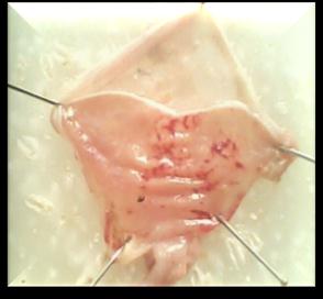
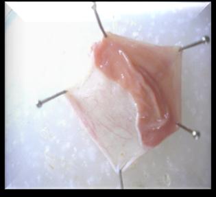
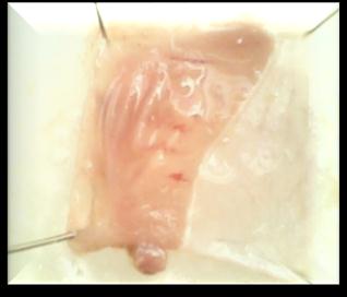
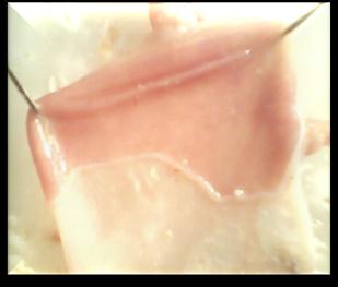
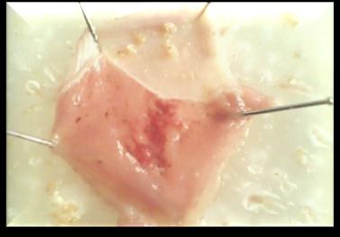
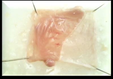
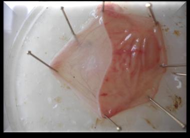
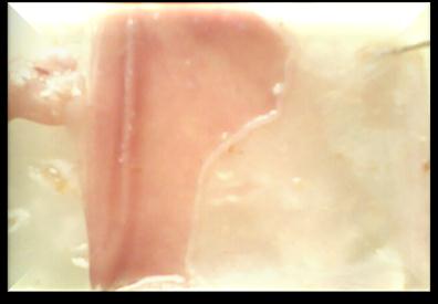
The images of the stomachs of the groups viz., Control, Standard and treatment groups (2 dose
levels of TE) in CRS (Figures 1-4) and aspirin-induced gastric lesion models (Figures 5-8) are
shown below.
Observations of ulcers in cold restraint model
Figure 1. Stress control group Figure 2. Standard (Cimetidine 100mg/kg) group
Figure 3. TE 400 mg/kg group Figure 4. TE 800 mg/kg group
Functional Foods in Health and Disease 2012, 2(3):48-61 Page 53 of 61
Observations of ulcers in aspirin-induced mucosal lesions
Figure 5. Toxicant (aspirin 200 mg/kg) Figure 6. Standard (omeprazole 20 mg/kg)
group group
Figure 7. TE 400 mg/kg group Figure 8. TE 800 mg/kg group
Marked ulcers along with hemorrhagic streaks and mucosal damage were seen in the images of
the toxicant group in the CRS (Figure 1) and the aspirin-induced gastric lesion model (Figure 5).
The images of the treatment groups showed signs of recovery from stress and ulcers and also
provided supportive evidence for the biochemical analysis (Figures 3-4 and 7-8).
Effect of TE on ulcer index and percentage lesion inhibition: The anti-ulcer activity of TE in
CRS and aspirin-induced gastric lesion models is reported in Tables 1 and 2 respectively. TE
treatment produced a significant reduction of the ulcer index (U.I.) (TE 400 mg/kg, P < 0.05; TE
800 and standard, P < 0.01) when compared with the stress control group in the CRS model and
the toxicant group in the aspirin induced ulcer model. The percentage lesion inhibition for TE
400 and TE 800 mg/kg was 23.13% and 38.48%, respectively in the CRS induced ulcer model
and 43.71% and 64.99%, respectively in the aspirin-induced gastric lesion model.
Table 1. Effect of TE on ulcer parameters in cold restraint model in rats
Treatment Groups/Doses
Mean Ulcer Score
Stress Control Group
Functional Foods in Health and Disease 2012, 2(3):48-61 Page 54 of 61
Test treatment group
Test treatment group
(Cimetidine 1.050±0.4463b
100mg/kg) group.
Values are mean ± SEM; N = 6 in each group P values : a < 0.05 when experimental groups compared with Stress Control b< 0.01 when experimental groups compared with Stress Control
Table 2. Effect of TE on ulcer parameters in aspirin induced ulcer model in rats.
Treatment Groups / Doses
Mean ulcer score
Ulcer Index
%Inhibition
Test treatment group
Test treatment group
(omeprazole 20 mg/kg).
Values are mean ± SEM; N = 6 in each group P values : a < 0.05 when experimental groups compared with Toxicant Control b< 0.01 when experimental groups compared with Toxicant Control
Effect of TE on the gastric tissue levels of GSH, TBARS and activities of antioxidant
enzymes SOD, CAT, GPx and GR in ulcer models: In order to determine the effect of TE on
oxidative stress induced in various ulcer models, the levels of GSH, TBARS and activities of
SOD, CAT, GPx and GR were measured in gastric tissue. Ulcer induction in both the models
was associated with marked reduction in the level of GSH, and activities of SOD, CAT, GPx &
GR, and an increase in the level of TBARS. Treatment with TE produced a significant increase
in the level of GSH and activities of SOD, CAT, GPx & GR, and a significant decrease in the
level of TBARS in the cold stress and aspirin models (Tables 3 and 4).
Table 3. Effect of TE on stomach GSH, TBARS, SOD, CAT, GPx and GR in cold restraint
model in rats
Biochemical
Group III
parameters
Cimetidine
(400 mg/kg)
800 mg/kg)
(100mg/kg)
1.68 ± 1.72 ± 0.08
Functional Foods in Health and Disease 2012, 2(3):48-61 Page 55 of 61
106.89 ± 17.17c
Values are mean ± SEM; N = 6 in each group
P values : a < 0.001 when Stress Control group compared with Normal Control group
b < 0.05, c < 0.01, d < 0.001 when experimental groups compared with Stress Control group
1 unit of CAT = µmol H2O2 consumed / min / mg protein
1 unit of GPX = µg GSH utilized / min / mg protein
1 unit of GR = nmol NADPH oxidized / min / mg protein
Table 4. Effect of TE on stomach GSH, TBARS, SOD, CAT, GPx and GR in aspirin
induced ulcer model in rats.
Biochemical
Group III
parameters
Toxicant
TE (400 mg/kg)
TE (800 mg/kg)
(omeprazole 20
protein) TBARS (nmol
31.59 ±0.75a 39.50 ±3.33b
Values are mean ± SEM; N = 6 in each group P values : a < 0.001 when Toxicant Control compared with Normal Control b < 0.05, c < 0.01 & d < 0.001 when experimental groups compared with Toxicant Control 1 unit of CAT = µmol H2O2 consumed / min / mg protein 1 unit of GPX = µg GSH utilized / min / mg protein 1 unit of GR = nmol NADPH oxidized / min / mg protein
DISCUSSION:
Tuber extract (TE) of Ipomoea batatas did not show any toxic or deleterious effects by oral route
up to 2000 mg/kg indicating low toxicity of the tuber extract at high doses. The LD50 value for
Functional Foods in Health and Disease 2012, 2(3):48-61 Page 56 of 61
the tuber extract by oral route could not be determined as no mortality was observed until a dose of 2000 mg/kg.
On preliminary phyto-chemical screening, the tuber extract revealed the presence of
phenolic compounds, polysaccharides, saponin glycosides and flavonoids as major compounds that might have contributed to its antioxidant activity. The present study was carried out to evaluate the ulcer protecting effect of Ipomoea batatas tuber extract in rats where gastric lesions were induced by cold stress and oral administration of aspirin.
Restraint stress has been one of the most popular stressors in experimental medicine. It
elicits the purest form of psychological frustration accompanied by vigorous struggling which means muscular exercise [25]. In the cold restraint model, stress in the control group clearly produced a mucosal damage characterized by multiple haemorrhage red bands of different sizes on the glandular stomach. Exposure of the animals to the cold restraint stress may have caused severe imbalance in the normal physiological conditions that might have resulted in a stressful condition leading to ulcers. Pre-treatment with test drugs (TE 400 and TE 800) produced a significant decrease in the intensity of gastric mucosal damage induced by the stress as compared with control. A significant increase in the ulcer index and mean score in the control group was observed, however both the parameters were significantly decreased in the TE 400 and TE 800 treated groups (Table 1). Further, examinations of the stomach images provide supportive evidence for the anti-ulcer activity of TE 400 and TE 800 treated groups which showed signs of recovery from the cold restraint stress induced ulcers (Figures 1-4). The reduction in the ulcer index and mean score may be attributed to the anti-ulcer activity of the tubers due to presence of antioxidants like anthocyanins, phenolic acids (eg, caffeic acid) and vitamin C.
Stress-induced ulcer is probably mediated by the release of histamine [26, 27]. It not only
increases gastric secretion, often called the „aggressive factor‟, but also causes disturbances of the gastric mucosal microcirculation and an abnormal motility, and reduces mucus production, known as the „defensive factor‟. Moreover, stress-induced ulcers in animal models may be partially or entirely prevented by vagotomy, since increased vagal activity has been suggested as the main factor in stress-induced ulceration. The vagus nerve stimulates stomach acid secretion via interaction of its chemical mediator (acetylcholine) with the muscarinic receptor. The activation of the muscarinic receptor gives rise to sequential events that result in increased gastric acid secretion. These receptors are located on the cell membranes of parietal cells and histamine secretory cells. Therefore, the increase in acid secretion is a consequence of acetylcholine action on the histamine cell and parietal cell activity. Stress-induced ulcers also involve damage by reactive oxygen species (ROS) apart from acid and pepsin related factors [25].
Exposure to cold restraint is a well known intensive stress response wherein both cold
exposure and immobilization individually and synergistically are responsible for the generation of reactive oxygen species (ROS) e.g. superoxide anion, hydrogen peroxide, hydroxyl radicals etc. that cause lipid peroxidation, especially in membranes and result in tissue injury. Elevation in the levels of end products of lipid peroxidation in stress control rat stomachs was observed. The increase in MDA levels in the stomach suggests enhanced peroxidation leading to tissue damage and failure of the antioxidant defense mechanisms to prevent formation of
Functional Foods in Health and Disease 2012, 2(3):48-61 Page 57 of 61
excessive free radicals. Pretreatment with TE 400 and TE 800 significantly reversed these changes (Table 3). Hence, it is likely that the mechanism of ant-ulceration of TE is due to its antioxidant effect.
Reduced glutathione is one of the most abundant non-enzymatic antioxidant bio-
molecules present in the tissues [28]. Its functions are removal of free oxygen species such as H2O2, superoxide anions & alkoxy radicals, maintenance of membrane protein thiols and to act as a substrate for GPx and glutathione S- transferase (GST) [29]. In the present study, stress enhanced the activities of GSH-related enzymes GPx and GST, thereby decreasing the GSH content, whereas treatment with TE reversed these effects (Table 3). It may be understood that the effect of TE may be due to increased synthesis of GSH or by stimulation of GR activity. Free radical scavenging enzymes such as SOD, CAT & GPx are known to be the first line cellular defense against oxidative damage, disposing O2 & H2O2 before their interaction to form the more harmful hydroxyl (OH·) radical [30]. In the present study SOD activity decreased significantly in the stress induced group of animals, which may be due to an excessive formation of superoxide anions (Table 3). These excessive superoxide anions might inactivate SOD and decrease its activity. The activities of the H2O2 scavenging enzymes CAT & GPx also decreased significantly in the stress induced group of animals. SOD is an important defense enzyme that catalyzes the dismutation of superoxide anions into O2 and H2O2 [31]. CAT is a hemeprotein that catalyzes the reduction of H2O2 to 2H2O and O2 and protects the tissue from highly reactive oxygen free radicals & hydroxyl radicals [32]. GPx, an enzyme with selenium, catalyzes the reduction of H2O2 and hydroperoxides to non-toxic products with the help of GSH [33]. GR is a cytosolic hepatic enzyme involved in the replenishment of glutathione stores by reduction of oxidized glutathione (GSSG), which is an end product of GPx activity [34]. In the stress induced group of animals, there was a marked reduction in GPx activity, leading to reduced availability of substrate for GR, thereby decreasing the activity of GR. Pretreatment with TE 400 and TE 800 significantly increased the activity of GR, thus accelerating the utilization of GSSG to form GSH and enhancing the detoxification of reactive metabolites by conjugation with GSH [35] (Table 3).
In this study SOD, CAT, GPx & GR activities were significantly elevated by administration of TE to treatment rats, suggesting that it has the ability to restore these enzymes‟ activities in stress control rats.
Functional Foods in Health and Disease 2012, 2(3):48-61 Page 58 of 61
In conclusion, the anti-ulcerative effect of TE against cold stress induced ulcer in rats
appears to be related to the inhibition of lipid peroxidative processes and to the prevention of GSH depletion. Furthermore, it may be stated that TE exerts its antioxidant activity by a dual action – by enzymatic as well as non-enzymatic pathways.
Nonsteroidal anti-inflammatory drugs (NSAIDs) such as aspirin have the ability to cause
gastroduodenal ulceration and this effect is related to the ability of these agents to suppress prostaglandin synthesis [36, 37, 38]. In the stomach, prostaglandins play a vital protective role, stimulating the secretion of bicarbonate and mucus, maintaining mucosal blood flow and regulating mucosal cell turnover and repair. Thus, the suppression of prostaglandin synthesis by NSAIDs results in increased susceptibility to mucosal injury and gastroduodenal ulceration [39]. One of the mechanisms by which aspirin damages the gastric mucosa is the increased production of NO due to the over expression of iNOS [40]. NO is a mediator not only of gastrointestinal mucosal defense [41], but also of its damage [42]. It has been shown that different concentrations of NO have completely opposite effects on the same tissue [43]. In general, the mucosal and endothelial NOS isoforms produce low amounts of NO. However, the high quantity of NO produced by iNOS damages the epithelium [43, 44]. The excessive release of NO from gastric epithelial cells induced by aspirin has been reported to exert detrimental effects [45].
In this study, exposure of the animals to aspirin may have caused severe ulcerogenic
effects, as aspirin is known to increase gastric acid secretion which is involved in the formation of aspirin-induced mucosal lesions.
However, gastric protection was observed by TE 400 and TE 800 in aspirin induced
gastric ulcers through parameters like mean score and ulcer index (Table 2). The gastro-protective activity of the TE 400 and TE 800 seems to be related to a reduction in the damage to the mucosa induced by free radicals and this activity may be due to its antioxidant action. These findings could be efficiently corroborated with the images of the aspirin induced animal group when compared with the TE 400 and TE 800 treated animal groups (figures 5-8).
Aspirin induced ulcer group of animals showed an increase in MDA level (Table 4) and a
decrease in GSH level (Table 4), as well as SOD, CAT, GPx and GR activities which was
reversed in TE 400 and TE 800 treatment groups (Table 4) probably by exhibiting a similar
mechanism as explained in cold restraint model.
CONCLUSION:
In conclusion, this study demonstrates that the tubers of Ipomoea batatas possess a potent ulcer
healing effect, which appears to be related to the free radical scavenging activity of the phyto
constituents, and their ability to inhibit lipid peroxidative processes. The present study, thus,
aims to highlight the health benefits of sweet potato, establish it as a potent "functional food"
and promote its use as a vegetable to enrich people‟s diets.
Competing interests: The authors declare that they have no competing interests.
Authors' contributions: Vandana Panda, conceived, designed, co-ordinated and supervised the
study and the writing of the manuscript. Madhav Sonkambale, initiated the study, carried out the
Functional Foods in Health and Disease 2012, 2(3):48-61 Page 59 of 61
experimental, performed statistical analysis and drafted the manuscript. All authors read and
approved the final manuscript.
Acknowledgement: We would like to thank Glenmark Pharmaceuticals Ltd. Mahape, Navi
Mumbai for providing animals for this research work.
REFERENCES:
1. Cullen D J, Hawkey G M, Greenwood D C:
Gut 1997, 41 (4): 459–62.
2. Mohan H: Textbook of Pathology, Jaypee Brothers Medical publishers (P) Ltd, New
Delhi; 2002, 4 : 526-535.
3. Berenguer L M, S'anchez A, Qu'ilez M: Protective and antioxidant activity of
Rhizophoro Mangle L. against NSAID‟S induced gastric ulcer. J. Ethanopharmacol 2006, 103:194-200.
4. Rang & Dale: The textbook of Pharmacology, An imprint of Elsevier. 2003, 5: 244-259. 5. Srikanta BM, Siddaraju MN, Dharmesh SM: A novel phenol-bound pectic
polysaccharide from Decalepis hamiltonii with multi-step ulcer preventive activity. World J Gastroenterol 2007, 13(39): 5196-207.
6. Maitya B, Chattopadhyay S: Natural Antiulcerogenic Agents: An Overview. Current
Bioactive Compounds 2008, 4: 225-44.
7. Samy RP, Gopalakrishnakone P: Current status of herbal and their future perspectives.
Nature Precedings 2007: 1-13.
8. Cordell GA, Plants in Drug Discoverv - Creating a New Vision. In: Tan BK, Bay BH,
Zhu YZ. Novel compounds from natural products in the new Millennium potential and challenges, National university of Singapore: World Scientific publishing; 2004. pp1-7.
9. Verma S, Singh SP: Current and future status of herbal medicines. Veterinary World
2008, 1(11): 347-50.
10. Scott G: Transforming traditional food crops: product development for roots and tubers.
In: Scott GJ, Wiersema S, Ferguson PI (Eds.), Product Development for Roots and Tuber Crops, vol. 1, Asia. International Potato Center (CIP), Lima, Peru; 1992:3–20.
11. Hayase F, Kato H: Antioxidative components of sweet potatoes. J. Nutritional Science
and Vitaminology 1984, 30: 37-46.
12. Kusano S, Abe H: Antidiabetic activity of white skinned sweet potato (Ipomoea batatas
L.) in obese Zucker fatty rats. Biological and Pharmaceutical Bulletin 2000, 23: 23-26.
13. Deore S L: Screening of anti stress properties of Chlorophytum borivilianum tuber.
Pharmacology online 2009, 1: 320-328.
14. Singh S, Majumdar D K: Evaluation of the gastric antiulcer activity of fixed oil of
Ocimum sanctum (Holy Basil). J. Ethnopharmacol 1999, 65:13–19.
15. Sairam K, Rao Ch.V, Dora Babu M, Vijay Kumar K, Agrawal VK, Goel RK:
Antiulcerogenic effect of methanolic extract of Emblica officinalis: an experimental study. J. of Ethnopharmacology 2002, 82: 1-9.
Functional Foods in Health and Disease 2012, 2(3):48-61 Page 60 of 61
16. Malairajan P, Gopalkrishnan G, Nrasimhan S, Jessi Kala Veni K, Kavimani S: Anti-ulcer
activity of crude alcoholic extract of Toona ciliata Roemer (heart wood). J. Ethanopharmacol 2007, 110: 348-357.
17. Ganguly A K, Bhatnagar O P: Effect of bilateral adrenalotomy on production of restraint
ulcers in stomach of albino rats. Canadian J. of Physiology and Pharmacology 1973, 51:748-750.
18. Ellman G L, Tissue sulphydryl groups. Arch Biochem Biophys 1959, 82: 70-77. 19. Sun M, Zigman S: An improved spectrophotometric assay for superoxide dismutase
based on epinephrine auto-oxidation. Anal. Biochem 1978, 90 (1): 81-89.
20. Clairborne A: Catalase Activity In: Greenwald RA CRS Handbook of Methods in
Oxygen Radical Research Boca Raton, CRS Press 1985: 283-284
21. Rotruck J T, Pope A L, Ganther H E, Hofeman D G, Hoekstra W G: Selenium:
Biochemical role as a component of Glutathione peroxidase. Science 1973, 179: 588-590.
22. Mohandas J, Marshal J J, Duggin G G, Harvasth J S,: Low activities of Glutathione-
related enzymes as factors in the genesis of urinary bladder cancer. Cancer Res. 1984, 44: 5086-91.
23. Lowry O H, Rosebrough N J, Farr A L, Randall R J: Protein Estimation with the Folin-
phenol reagent. J. Biol. Chem. 1951, 193: 265-275.
24. Ohkawa H, Oshishi N, Yagi K: Assay of lipid peroxidation in animal tissue by
thiobarbituric acid reaction. Anal. Biochem. 1979, 95:351-58.
25. Bhattacharya A, Ghosal S, Bhattacharya S K: Anti-oxidant effect of Withania somnifera
glycowithanolides in chronic foot shock stress-induced perturbations of oxidative free radical scavenging enzymes and lipid peroxidation in rat frontal cortex and striatum. J. Ethnopharmacol 2001, 74: 1–6.
26. Michael N P, Charles T R: Stressful life events, acid hypersecretion and ulcer disease.
Gastroenterology 1983, 84:114–119.
27. Brodie D A Q, Hanson H M: A study of the factors involved in the production of gastric
ulcer by the restraint technique. Gastroenterology 1960, 38:353–360.
28. Meister A: New aspects of glutathione biochemistry and transport selective alteration of
glutathione metabolism. Nutr. Rev. 1984, 42: 397-400.
29. Townsend DM, Tew KD, Tapiero H: The importance of glutathione in human disease.
Biomed. Pharmacother. 2003, 57:144-155.
30. Lil JL, Stantman FW, Lardy HA. Antioxidant enzyme systems in rat liver and skeletal
muscle. Arch. Biochem. Biophys.1988; 263:150-160.
31. Bannister J, Bannister W: Aspects of the structure, function and application of superoxide
dismutase. CRC Crit Rev Biochem 1987, 22 (2): 111-80.
32. Zamocky M, Koller F: Understanding the structure and function of catalases clues from
molecular evolution and in vitro mutagenesis. Prog Biophy. Mol Bio 1999, 72 (1): 1966
33. Bruce A, Freeman D, James C: Biology of disease-free radicals and tissue injury. Lab.
Invest. 1982, 47: 412-426.
34. Manneersk B: The enzymes of glutathione metabolism: an overview. Biochem Soc Trans
1987, 15 (4):717- 8.
Functional Foods in Health and Disease 2012, 2(3):48-61 Page 61 of 61
35. Naik S, Panda V: Hepatoprotective effect of Ginkgoselect Phytosome® in rifampicin
induced liver injurym in rats: Evidence of antioxidant activity, Fitoterapia 2008, 79 (6):439- 445.
36. Takeuchi K, Ueki S, Tanaka H: Endogenous prostaglandins in gastric alkaline response
in the rat stomach after damage. American J. Physiology 1986, 250: G842–G849.
37. Lichtenberger L M, Romero J J, Dial E J: Surface phospholipids in gastric injury and
protection when a selective cyclooxygenase-2 inhibitor (Coxib) is used in combination with aspirin. British J. Pharmacology 2007, 150: 913–919.
38. Wang GZ, Huang GP, Yin GL, Zhou G, Guo, CJ, Xie CG: Aspirin can elicit the
recurrence of gastric ulcer induced with acetic acid in rats. Cellular Physiology and Biochemistry 2007, 20: 205–212.
39. Deore A, Sapakal V, Dashputre N, Naikwade N: Antiulcer activity of Garcinia indica
linn fruit rinds. J. Applied Pharmaceutical Science 2011, 1 (5): 151-154.
40. Konturek PC, Kania J, Hahn EG, Konturek JW: Ascorbic acid attenuates aspirin-induced
gastric damage: role of inducible nitric oxide synthase. J. Physiology and Pharmacology
2006, 57:125–136.
41. Calatayud S, Barrachina D, Espluges JV: Nitric oxide: relation to integrity, injury, and
healing of the gastric mucosa. Microsc Res Tech 2001, 53: 325–335.
42. Muscara MN, Wallace JL. Nitric oxide. V: Therapeutic potential of nitric oxide donors
and inhibitors. Am. J. Physiol 1999, 276: G1313–G1316.
43. Wallace JL, Miller MJ: Nitric oxide in mucosal defense: a little goes a long way.
Gastroenterol 2000, 119: 512–520.
44. Piotrowski J, Slomiany A, Slomiany BL: Activation of apoptotic caspase-3 and nitric
oxide synthase-2 in gastric mucosal injury induced by indomethacin. Scandinavian J.
Gastroenterol 1999, 34: 129–134.
45. Whittle BJ: Gastrointestinal effects of nonsteroidal anti-inflammatory drugs.
Fundamental & Clinical Pharmacol 2003, 7: 301–313.
Source: http://www.functionalfoodscenter.net/files/50426782.pdf
Investigación Captopril por vía oral y sublingual en pacientes con urgencia hipertensiva Captopril orally and sublingually in patients with hypertensive urgency 1Arquimides Mackelin Ortega Vázquez, 2Norma Elena Corona Amador. 1Adscripción: UMF 56 León; Guanajuato. 2Adscripción: HGZ/UMF 21 León; Guanajuato. Resumen Introducción La Urgencia hipertensiva se presenta generalmente en enfermos con hipertensión arterial sistémica. El 1% de los pacientes hipertensos tendrán crisis hipertensiva de la cual el 76% será Urgencia hipertensiva y 24% emergencia ( Papaduopulos D,2010). Objetivo Evaluar si el Captopril sublingual es mejor para el control de la urgencia hipertensiva que el captopril vía ora en los pacientes atendidos en urgencias de la UMF 56 de Julio a Diciembre del 2013. Material y métodos Se realizó un estudio cuasi experimental, el tamaño de la muestra se calculó de la diferencia entre dos proporciones, con un nivel de confianza del 95%, con 40 pacientes en cada uno de los grupos (sublingual Vs oral); por casos consecutivos y de forma aleatoria se asignó al grupo oral o sublingual, en ambos casos se administró molida la tableta de captopril. La población de estudio fueron los derechohabientes que acudieron al servicio de urgencias de UMF 56 de Julio a Diciembre del 2013, y que cumplieron los criterios de diagnóstico de urgencia hipertensiva. Resultados Captopril molido sublingual mostro mayor rapidez en cuanto el control de la Tensión Arterial Media ≤ 100mmHg y disminución TAM ≥ 20%, Comparando los promedios de control de las TAM de ambos grupos encontramos una diferencia estadísticamente significativa de P= 0.0035. Conclusión Observamos que el 16.25% de la muestra desconocía ser hipertenso. El 82.35% (42 pacientes) de los pacientes descontrolados son manejados con un anti-hipertensivo. Encontramos una relación directamente proporcional del índice de masa corporal y al grado de descontrol de hipertensión a su ingreso.
18 al 20 de febrero de 2009 3er Foro Latinoamericano sobre Higiene íntima Femenina Actualización en patología vulvar y tracto urinario Del 18 al 20 de febrero de 2009 se realizó en la Ciudad de Varadero, Cuba, el 3er Foro Latinoamericano sobre Higiene Íntima Femenina. En esta ocasión, el evento estuvo dirigido a la actualización en patología vulvar y tracto urinario. Diversos especialistas de países latinoamericanos comentaron sus experiencias con el objetivo de actualizar al médico ginecólogo en la etiología, el diagnóstico y el tratamiento de las distintas afecciones vulvares y vaginales. Se contó con la presencia del Dr. Jaime Piquero Casals (Venezuela), quien habló sobre los aspectos clínicos de las vulvitis frecuentes y de la problemática de la infección vulvar por HPV. Las distrofias vulvares, especialmente el liquen escleroso y el liquen simple crónico, fueron comentadas por la Dra. Lina María Figueira (Venezuela). El Dr. Wel ington Aguirre (Ecuador) se refirió a los trastornos genitourinarios en la menopausia y su abordaje farmacológico. El Dr. Alejandro Paradas (República Dominicana) disertó acerca de la protección y la prevención de las infecciones vaginales. Finalmente, el Dr. Santiago Herrán (Colombia) expuso los resultados del primer estudio epidemiológico latinoamericano sobre hábitos de higiene íntima femenina y su relación con la vaginosis bacteriana en mujeres latinoamericanas, inquietud que tuvo su origen en el foro predecesor realizado en 2008 en Panamá.Surge como principal conclusión de este encuentro que la adopción de hábitos de higiene íntima femenina adecuados es una medida esencial en la prevención de afecciones genitourinarias tanto de origen infeccioso como no infeccioso.


